Summary
Description
Osteoblastoma is benign tumor which creates bone and osteoid. Lesions are commonly found on the dorsal aspect of the vertebrae or long bones.
People and Age
Most commonly occurs in young adults. with a peak age of incidence in the 20's.
Symptoms and Presentation
Symptoms include sustained pain, swelling and tenderness. Spinal tumors can cause scoliosis and neurological symptoms.
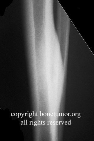
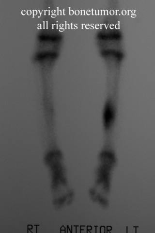
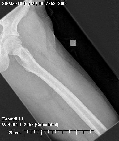
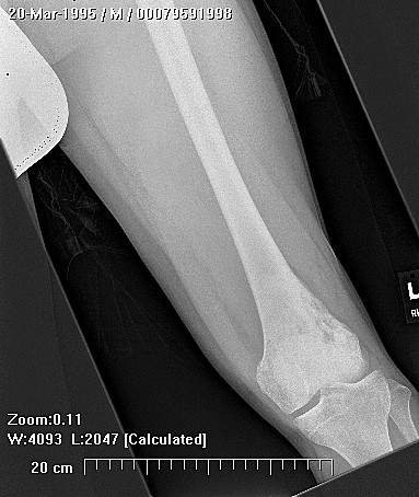
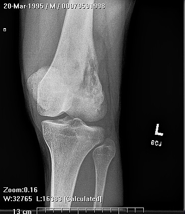
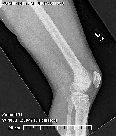
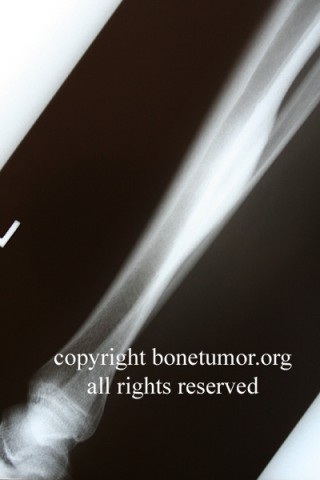
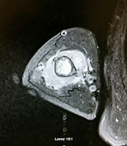
Brief description of the xray
The lesion appears as radio-lucent defect with central density. Tumor also displays increased isotope uptake.
Tumor Name
Tumor Type
Benign or Malignant
Body region
Most Common Bones
Periosteal reaction
Tumor behavior
Tumor density
Complete Information on this Tumor
Introduction and Definition
Osteoblastoma is a solitary, benign bone-forming tumor that occurs in the spine and long bones of young adults. Osteoblastoma and osteoid osteoma are histologically very similar, yet these two tumors are very different in their presentation, localization, radiographic appearance, treatment, and potential for recurrence.
Incidence and Demographics
The tumor most commonly occurs in the dorsal aspect of vertebrae, the metaphysis or diaphysis of long bones, and rarely in the pelvis.In the spine, the tumor is usually located in the posterior processes while the vertebral bodies are spared. Also, though tumor frequency is lower in the thoracic region of the spine, it has greater and equal occurrence in the cervical and lumbar regions.
Osteoblastoma predominantly affects young adults. The peak age of occurrence is approximately age twenty, though the tumor may present as early as age ten to as late as age sixty.
Osteoblastoma predominantly affects young adults. The peak age of occurrence is approximately age twenty, though the tumor may present as early as age ten to as late as age sixty.
Symptoms and Presentation
Common symptoms are pain of long duration, swelling and tenderness. Tumors of the spine can cause scoliosis and neurological symptoms. The lesion may clinically present with myleopathic and/or radicular symptoms.
X-Ray Appearance and Advanced Imaging Findings
On x-ray, osteoblastomas appear as a radio-lucent defect with a central density due to ossification. The lesion is well circumscribed and may have a surrounding sclerosis. The tumor demonstrates increased isotope uptake on bone scan.
Differential Diagnosis
The differential diagnosis of osteoblastoma includes osteoid osteoma, osteosarcoma, giant cell tumor and aneurysmal bone cyst.
Histopathology findings
On gross examination, osteoblastomas are red to tan in color with hemorrhagic areas. The compact tissue is granular, friable and gritty. Hyperemia is particularly evident in the spongy bone of vertebrae, ribs and the pelvis.
The classic microscopic finding of osteoblastoma is irregular spicules of mineralized bone and eosinophilic osteoid rimmed by osteoblasts. The vascular stroma is characterized by pleomorphic spindle cells. The tumor cells differentiate into osteoblasts which make varying amounts of osteoid and woven bone. Cartilage production is a very rare finding in an osteoblastoma and should raise the suspicion of osteosarcoma.
The classic microscopic finding of osteoblastoma is irregular spicules of mineralized bone and eosinophilic osteoid rimmed by osteoblasts. The vascular stroma is characterized by pleomorphic spindle cells. The tumor cells differentiate into osteoblasts which make varying amounts of osteoid and woven bone. Cartilage production is a very rare finding in an osteoblastoma and should raise the suspicion of osteosarcoma.
Treatment Options for this Tumor
Usually,a biopsy is performed to confirm the diagnosis. Surgical resection by curettage, intralesional excision or en-bloc excision are all treatment options depending on the site. Cryosurgery, radiation and chemotherapy may have a role in aggressive and surgically unresectable lesions of the spine.
Preferred Margin for this Tumor
Margins should be a s wide as possible without functional sacrifice.
Special and Unusual Features
Osteoblastoma vs. Osteoid Osteoma
inconsistent pain vs.persistent, nocturnal pain
irregular tissue pattern vs. regular pattern
>2 cm vs. < 1 cm
Sporadic reports of malignant sarcomas arising in osteoblastoma have been published. In addition, multiple authors have described a subset of these tumors that behaves in a much more locally aggressive fashion. These tumors have been found to be larger and occur in slightly older individuals. Microscopically, these tumors may have a distinct appearance, including epithelioid features and larger osteoblasts with abundant eosinophilic cytoplasm and vesicular nuclei. These tumors have been variously termed "aggressive osteoblastoma" or "malignant osteoblastoma". The radiographic and pathologic features of these tumors overlap with osteosarcoma.
Suggested Reading and Reference
Sources
Bullough, Peter, Orthopaedic Pathologv (third edition), Times Mirror International Publishers Limited: London, 1997.
Gitelis S., R. Wilkins and EU Conrad, Benign Bone Tumors. Instructional Course Lectures, 45:425-46, 1991.
Huvos, Andrew, Bone Tumors: Diagnosis. Treatment and Prognosis, W.B. Saunders Co., 1991.
Ruggieri, P., RA McLeod, KK Unni and FH Sim, Osteoblastoma, Orthopedics, 19(7):621-4, July 1996.