Summary
Description
Adamantinoma of the long bones, or extragnathic adamantinoma, is an extremely rare, slow growing, low-grade malignant tumor of epithelial origin, that occurs almost exclusively in the tibia and the fibula.
People and Age
The tumor usually occurs in the second to fifth decade of life.
Symptoms and Presentation
The patient usually has swelling that may be painful. The duration of symptoms can vary from a few weeks to years.
Brief description of the xray
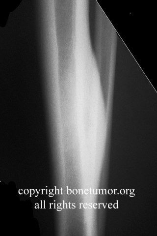
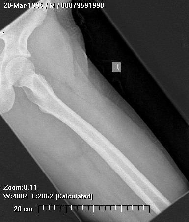
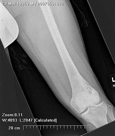
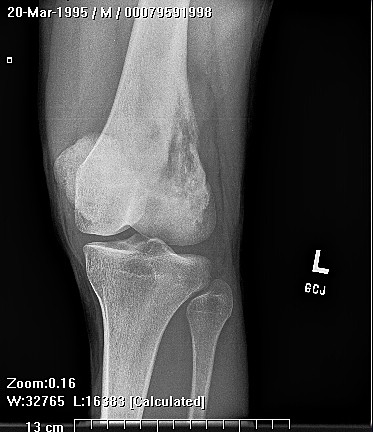
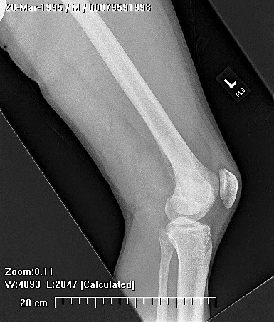
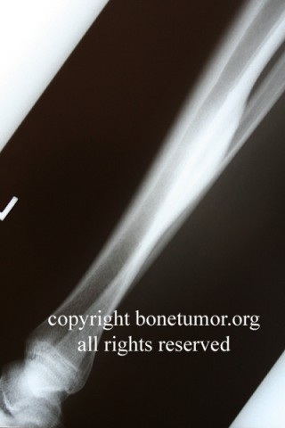
Adamantinoma appears as an eccentric, well-circumscribed, and lytic lesion on plain x-ray.
Brief desc of tx
Adamantinoma is treated by wide surgical excision
Complete Information on this Tumor
Introduction and Definition
Adamantinoma of the long bones, or extragnathic adamantinoma, is an extremely rare, low-grade malignant tumor of epithelial origin. It is not related to adamantinoma or ameloblastoma of the mandible and maxilla which is derived from Rathke's pouch. Adamantinoma is a locally aggressive osteolytic tumor
Incidence and Demographics
The tumor usually occurs in the second to fifth decade of life but may affect patients from ages 3 to 73. Most patients are male.
Symptoms and Presentation
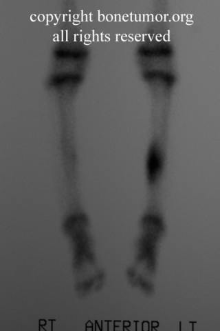
The site 90% of the time in the diaphysis of the tibia with the remaining lesions found in the fibula and long tubular bones. There is often a history of trauma associated with adamantinoma but its role in the development of this lesion remains unclear. The patient usually has swelling that may be painful. The duration of symptoms can vary from a few weeks to years.
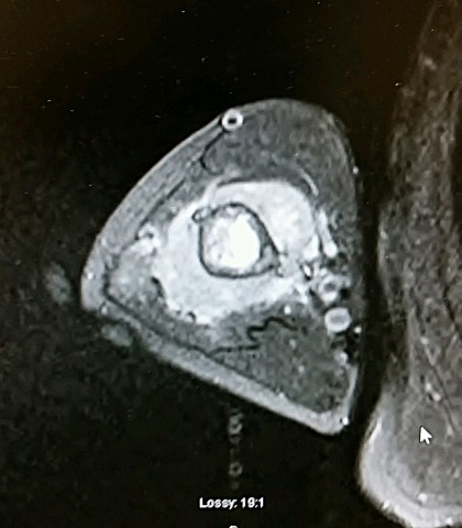
X-Ray Appearance and Advanced Imaging Findings
Adamantinoma appears as an eccentric, well-circumscribed, and lytic lesion on plain x-ray. The anterior cortex of the tibia is by far the most common location. The lesion usually has several lytic defects separated by sclerotic bone which gives a "soap-bubble" appearance. There is cortical thinning but little periosteal reaction. The lesion may break through the cortex and extend into soft tissue. There may be multiple adjacent lesions with normal intervening bone. MRI helps demonstrate the intraosseus and extraosseous involvement. The differential diagnosis radiologically includes osteofibrous dysplasia, fibrous dysplasia, ABC, chondromyxoid fibroma and chondrosarcoma
Differential Diagnosis
The differential diagnosis radiologically includes osteofibrous dysplasia, fibrous dysplasia, ABC, chondromyxoid fibroma and chondrosarcoma .
Preferred Biopsy Technique for this Tumor
Open biopsy
Histopathology findings
On gross examination, adamantinoma is well demarcated and lobulated. The gray or white tumor is rubbery and may have focal areas of hemorrhage and necrosis. Bone spicules and cysts filled with blood or straw-colored fluid may also be present.Adamantinoma is a biphasic tumor with islands of epithelioid cells surrounded by a bland reactive fibrous stroma. The stroma consists of spindle shaped collagen producing cells. The nests of malignant cells are columnar and have peripheral palisading. Squamous differentiation and keratin production are rare. The tumor is positive on immunohistochemical staining with keratin antibody. The epithelial origin is confirmed when basal membranes, desmosomes and ton filaments are seen under the electron microscope.
Treatment Options for this Tumor
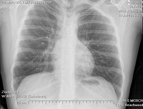
Adamantinoma is treated by wide surgical excision. This tumor is insensitive to radiation and may metastasize to lungs, lymph nodes and abdominal organs by both hematogenous and lymphatic routes. Chemotherapy is not used.
Preferred Margin for this Tumor
wide
Outcomes of Treatment and Prognosis
In 20% of cases there are metastases late in the course of the disease.
Special and Unusual Features
Osteofibrous dysplasia, or ossifying fibroma, is another lesion with a striking predilection for the tibia that has a well documented association with adamantinoma and may be a benign precursor to it.
Suggested Reading and Reference
Conway, WF and CW Hayes, Miscellaneous Lesions of Bone, Radiologic Clinics of North America, 31(2):339357, March, 1993.
Bulloughs, Andrew, Orthopaedic Pathologv (third edition), Times Mirror International Publishers Limited, London, 1997
Huvos, Andew. Bone Tumors- Diagnosis. Treatment and Prognosis, W.B.Saunders, Co., 1991.
Fletcher, Christopher. Diagnostic Histopathology of Tumors, Churchill Livingstone, 1995.
Van Der Woude, HJ AJR Am J Roentgenol. 2004 Dec;183(6):1737-44.