Summary
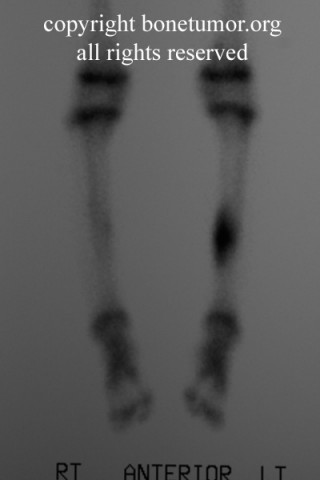
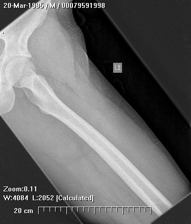
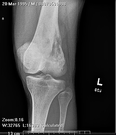
Epithelioid hemangioendothelioma (EHE) is a rare, well-differentiated endothelial tumor with a wide spectrum of behavior. It occurs most commonly in the calvarium, spine, femur, tibia and feet of adults.
Complete Information on this Tumor
Epithelioid hemangioendothelioma (EHE) is a rare, well-differentiated endothelial tumor with a wide spectrum of behavior. The term was designed to describe tumors that had an appearance in between hemangiomas and sarcomas. Epithelioid hemangioendothelioma has several synonyms: low grade anaplastic angiosarcoma, cellular hemangioma, histiocytoid hemangioma and angioendothelioma. It represents 1% of all vascular neoplasms and is locally aggressive.
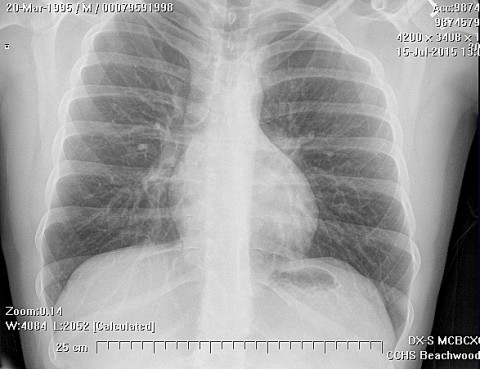
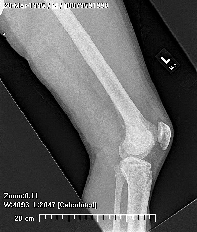
Clinically, epithelioid hemangioendotheliomas presents with pain and swelling. If present in the spine, a lesion may cause radicular symptoms or paraplegia.
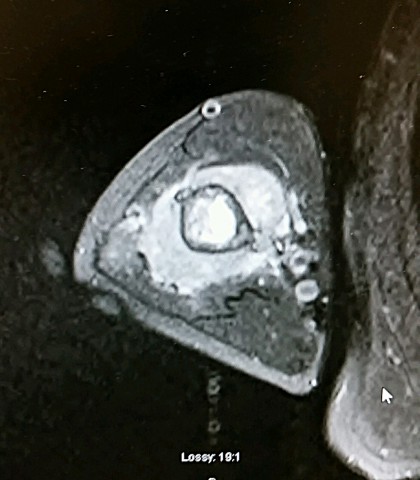
Microscopically, epithelioid hemangioendotheliomas appear as irregular anastomosing vascular channels. The channels are lined by plump endothelial cells without pleomorphism or mitotic activity. Both the background stroma and the cells lining the vascular channels stain positive for reticulum. The epithelioid histological subtype of epithelioid hemangioendothelioma has epithelial like cells lining the vascular channels. The spindle cell variant has spindle cells separating the vessels. Various criteria are used to determine if the lesion is benign or malignant including the number of mitoses, hyperchromatic nuclei, pleomorphism and nucleus to cell ratio.
Huvos, Andrew. Bone Tumors: Diagnosis. Treatment and Prognosis, W.B. Saunders, Co., 1991.
Parry, Bryan, A 23-Year-Old Man With Pain and Swelling in His Left Thumb, The American Journal of Orthopedics, p. 725-728, September, 1995.