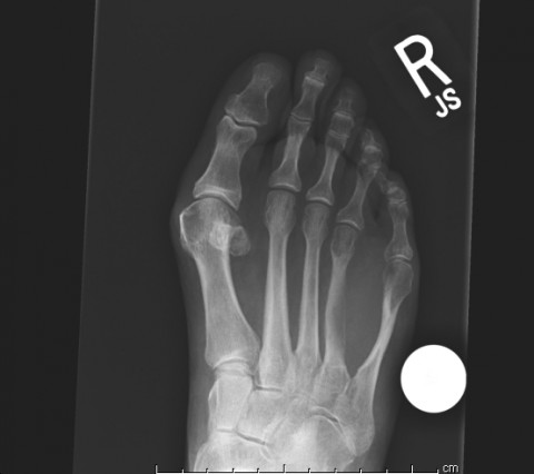
A very large forefoot tumor in a 70 year old woman Case ID Number 20150430VL Image Types Main Image Image modality Xray Tumor Name unknown tumor - soft tissue Example Image yes Tumor Type unknown tumor - soft tissue Benign or Malignant unknown Body region ankle and foot, leg
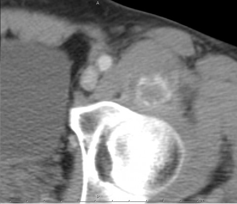
In the hospital 5 days for hip pain, but he went home because it got worse Case ID Number GW20140624 Image Types Main Image Image modality CT Tumor Name unknown tumor - soft tissue Example Image yes Tumor Type unknown tumor - soft tissue Benign or Malignant unknown Body region pelvis - entire
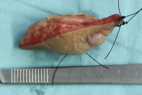
Nicaragua 2013 - Case 3 - Patient Y.M.H. - Female 14 left medial ankle soft tissue mass Case ID Number 2013Nica3 Image Types Auxiliary Image Image modality gross photo Tumor Name unknown tumor - soft tissue Example Image yes Tumor Type unknown tumor - soft tissue Benign or Malignant unknown Body region ankle and foot, leg
Nicaragua 2013 - Case 6 - Patient V.V.E - 49 Female Multifocal soft tissue lesion carpal tunnel 5th finger flexor sheath Read more about Nicaragua 2013 - Case 6 - Patient V.V.E - 49 Female Multifocal soft tissue lesion carpal tunnel 5th finger flexor sheath
Nicaragua 2013 - Case 3 - Patient Y.M.H. - Female 14 left medial ankle soft tissue mass Read more about Nicaragua 2013 - Case 3 - Patient Y.M.H. - Female 14 left medial ankle soft tissue mass
Nicaragua 2013 - Case 2 - Patient C.J.C. - Male 15 right distal quads mass Read more about Nicaragua 2013 - Case 2 - Patient C.J.C. - Male 15 right distal quads mass
Nicaragua 2013 - Case 1 - Patient G.J.R.C. Male 65 left posterior thigh mass Read more about Nicaragua 2013 - Case 1 - Patient G.J.R.C. Male 65 left posterior thigh mass
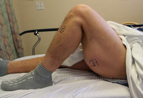
A man with multiple skin tumors and a thigh mass Case ID Number 20131212TM Image Types Auxiliary Image Image modality clinical photo Tumor Name unknown tumor - soft tissue Example Image yes Tumor Type unknown tumor - soft tissue Benign or Malignant unknown Body region knee, thigh, distal femur, proximal tibia
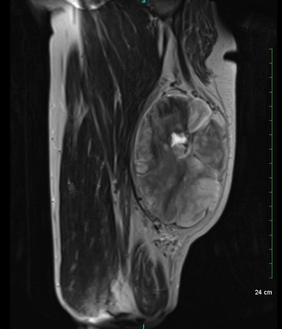
A man with multiple skin tumors and a thigh mass Case ID Number 20131212TM Image Types Auxiliary Image Image modality MRI Tumor Name unknown tumor - soft tissue Example Image yes Tumor Type unknown tumor - soft tissue Benign or Malignant unknown Body region knee, thigh, distal femur, proximal tibia
A man with multiple skin tumors and a thigh mass Case ID Number 20131212TM Image Types Main Image Image modality MRI Tumor Name unknown tumor - soft tissue Example Image yes Tumor Type unknown tumor - soft tissue Benign or Malignant unknown Body region knee, thigh, distal femur, proximal tibia