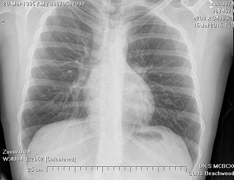
In the hospital 5 days for hip pain, but he went home because it got worse Case ID Number GW20140624 Image Types Main Image Image modality CT Tumor Name unknown tumor - soft tissue Example Image yes Tumor Type unknown tumor - soft tissue Benign or Malignant unknown Body region pelvis - entire
In the hospital 5 days for hip pain, but he went home because it got worse Read more about In the hospital 5 days for hip pain, but he went home because it got worse
Erdheim–Chester disease (ECD) Secret Tumor Name Erdheim–Chester disease Image Types Auxiliary Image Image modality Xray Tumor Name Erdheim–Chester disease Example Image yes Benign or Malignant Benign Body region pelvis - entire Bone name ilium Tumor behavior latent
Erdheim–Chester disease (ECD) Secret Tumor Name Erdheim–Chester disease Image Types Main Image Image modality Xray Tumor Name Erdheim–Chester disease Example Image yes Benign or Malignant Benign Body region pelvis - entire Bone name ilium Tumor behavior latent
Erdheim–Chester disease Secret Tumor Name Erdheim–Chester disease Image Types Main Image Image modality clinical photo Tumor Name Erdheim–Chester disease Example Image yes Benign or Malignant Benign Body region pelvis - entire Bone name ilium
A 95 year old lady with increasing pain and swelling in her thigh Case ID Number 20120918SF Image Types Auxiliary Image Image modality CT Tumor Name Lymphoma of bone Example Image no Benign or Malignant Benign Body region pelvis - entire position within the bone central
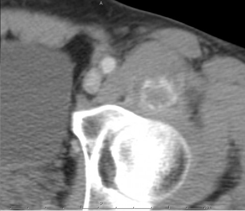
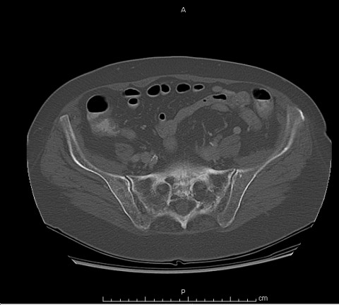
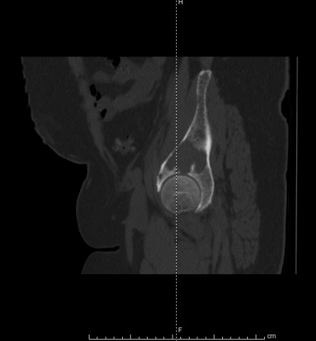
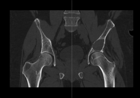
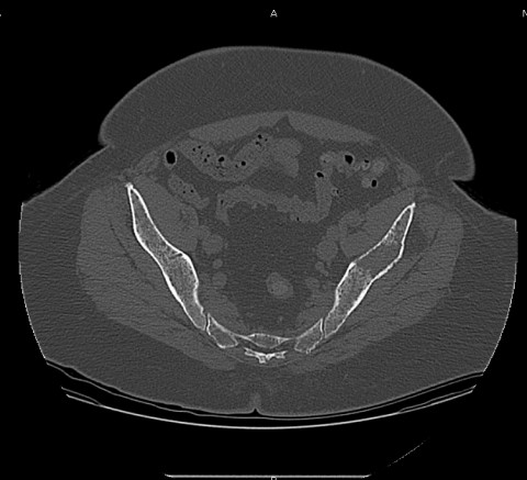
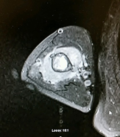
52 year old man with left hip pain Case ID Number 20120907BT Image Types Auxiliary Image Image modality CT Tumor Name Multiple myeloma Example Image yes Benign or Malignant Malignant Body region pelvis - entire Bone name ilium periosteal reaction absent position within the bone central Tumor behavior aggressive Tumor density lytic
52 year old man with left hip pain Case ID Number 20120907BT Image Types Auxiliary Image Image modality CT Tumor Name Multiple myeloma Example Image yes Tumor Type Plasma cell tumors Benign or Malignant Malignant Body region pelvis - entire Bone name ilium periosteal reaction absent position within the bone central Tumor behavior aggressive Tumor density lytic
52 year old man with left hip pain Case ID Number 20120907BT Image Types Auxiliary Image Image modality CT Tumor Name Multiple myeloma Example Image yes Tumor Type Plasma cell tumors Benign or Malignant Malignant Body region pelvis - entire Bone name ilium periosteal reaction absent position within the bone central Tumor behavior aggressive Tumor density lytic
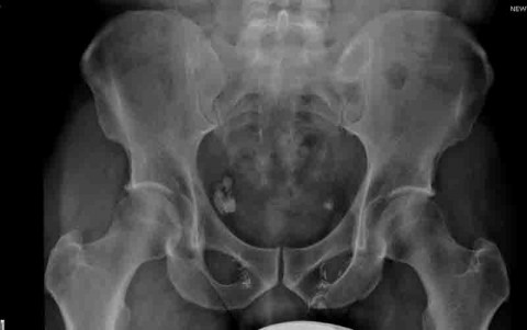
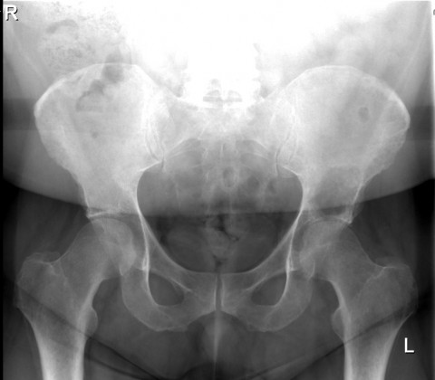
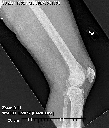
52 year old man with left hip pain Case ID Number 20120907BT Image Types Main Image Image modality Xray Tumor Name Multiple myeloma Example Image no Tumor Type Plasma cell tumors Benign or Malignant Malignant Body region pelvis - entire position within the bone central Tumor density lytic