McCune-Albright syndrome (MAS)

Summary
Description

McCune-Albright syndrome (MAS) is a rare, genetic, non-inherited condition that causes bone tumors, bone deformity and fractures. The manifestations include polyostotic fibrous dysplasia, café au lait spots, and precocious puberty.

Symptoms and Presentation
The clinical findings vary widely. Some patients have bone pain while others have no symptoms.
Brief description of the xray
On radiographs, the affected bones have multiple, expansile, mostly lucent lesions which contained a fine matrix described as " ground glass.
Brief desc of tx
Treatment of weak bones may require surgery. Medical treatments with bisphosphonates are helpful.
Benign or Malignant
Most Common Bones
Location in bone
Periosteal reaction
Position within the bone
Tumor behavior
Complete Information on this Tumor
Introduction and Definition

McCune-Albright syndrome (MAS) is a rare condition that results from a spontaneous, non-inherited mutation in the Gs gene. The manifestations include polyostotic fibrous dysplasia, bone fractures, café au lait (hyperpigmented) skin spots, precocious onset of puberty, and variable hyperactivity of the endocrine glands.

Symptoms and Presentation

Mutation of the GS gene in chromosome 20q13 occurs early in development, and results in a mosaic of abnormal and mutated cells. The manifestations of MAS in each individual depend upon the extent and distribution of abnormal cells. Abnormal and prolonged activation of multiple peripheral endocrine glands occurs even while the necessary stimulatory pituitary hormones may be absent. Precocious puberty, with onset of breast development, pubic hair, and the onset of menses as early as the first few months of life may occur in females. Other manifestations include acromegaly, hyperthyroidism, hyperprolactinemia, and others.

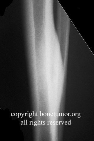
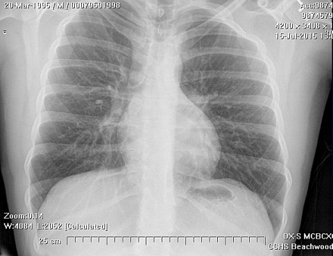
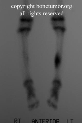
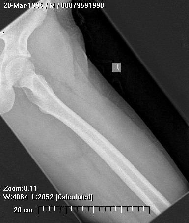
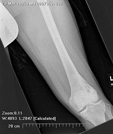
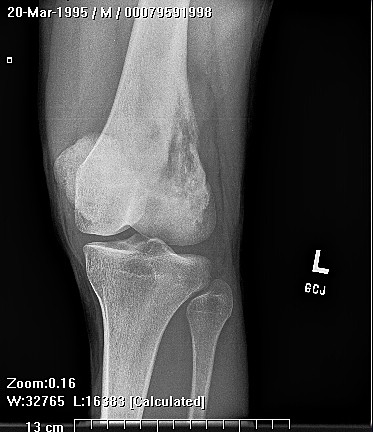
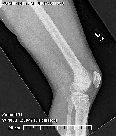
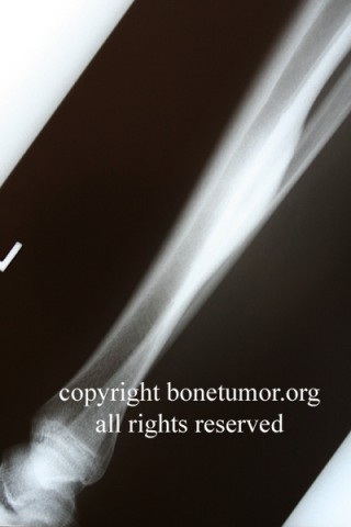
Frequently involved bones include the femur, the tibia, the facial skeleton, and the ribs. Bone fragility and associated fractures are common, and weight-bearing bones may suffer multiple fractures. In the proximal femurs, multiple successive cortical microfractures may result in characteristic bowing of the proximal end of the bone into a "shepherd's crook" deformity.

X-Ray Appearance and Advanced Imaging Findings
On radiographs, the affected bones have multiple, expansile, mostly lucent lesions which contained a fine matrix described as " ground glass." The normal trabecular pattern is absent. The tumors are a radiolucent area with a sclerotic rim, and may slightly expand or thin the nearby bone cortex. The matrix of the lesion has a "milky" or "ground glass" appearance due to the very fine bony trabeculae contained within the tumor. On bone scan, fibrous dysplasia lesions typically demonstrate increased uptake. However, 10% to 15% of lesions do not have increased radiotracer uptake and appear "cold" on bone scans.
Histopathology findings
The pathological findings in the involved bones include classic features of fibrous dysplasia. On microscopic analysis, there is fibrous connective tissue containing immature trabeculae of a woven, non-lamellar bone. These immature trabeculae may have a characteristic "Chinese letter" or "alphabet soup" appearance, resembling partially formed letters or symbols.
Treatment Options for this Tumor
Treatment depends on the manifestations and extent of the disease. Bone fracture lead to the need for orthopedic stabilization and joint replacement due to periarticular fracures. Pamidronate treatment of patient's with polyostotic fibrous dysplasia has shown encouraging results. Pamidronate seems to reduce the fracture rate and reduce the bone pain associated with the lesions. The benefit of pamidronate in polyostotic fibrous dysplasia has been conclusively demonstrated. Bone pain, fracture risks, bone density, and metabolic indices of bone turnover show favorable response to treatment. However, some authors have reported that in developing children, dysplastic lesions in long bones continued to undergo expansion despite pamidronate treatment. For developing children, the authors of this website recommend early and aggressive intervention with pamidronate, frequent follow-up, and orthopedic stabilization of at-risk long bones, using appropriate expandable or non-expandable intramedullary devices. The femoral neck and proximal femur are at particularly high risk, and early prophylactic intramedullary rodding with a cephalomedullary device is recommended, before the microfractures and deformity begin to develop.
Outcomes of Treatment and Prognosis
The general overall prognosis for patients with this syndrome is good, but the extent of involvement may lead to severe dysfunction of affected organs, including the hepatobiliary system, cardiopulmonary system, and virtually any affected organ, leading to risk factors for early death.
Special and Unusual Features
The risk of malignant degeneration in fibrous dysplasia is a special cause for concern in patients with McCune-Albright syndrome. In the largest available review, Ruggieri examined 1122 cases from the Mayo Clinic amongst which he found 28 cases uf sarcoma. In these 28 cases, 19 cases occurred in modest fibrous dysplasia and 9 cases occurred in polyostotic disease. Only one case occurred in McCune-Albrights syndrome. The sarcomas were classified as osteosarcoma in 19 cases, fibrosarcoma in 5 cases, chondrosarcoma in 3 cases, and malignant fibrous histiocytoma in one case. 46% of the cases had received radiation therapy before the sarcoma developed. Most occurred in a cranial facial bones or the proximal femur.

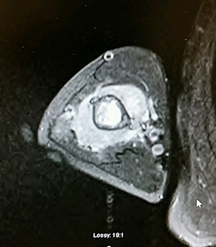
Clinical signs of sarcomatous degeneration include pain and swelling at the site of the lesion. The most reliable radiographic sign is extension of the lesion through the cortex into the surrounding soft tissues. Since osteosarcoma is the most common histologic type of sarcoma that may occur, bone formation within the soft tissue mass would be a particularly worrisome sign.