Summary
Description
Osteoid Osteoma is a benign bone lesion with a nidus of less than 2 cm surrounded by a zone of reactive bone. It occurs most commonly in the long bones, especially the femur, and the posterior spine.
People and Age
The tumor presents by the twenties, with a peak age in the early twenties. The male to female ratio is 2:1.
Symptoms and Presentation
Symptoms include dull pain, increase in skin temperature, increased sweating and tenderness.
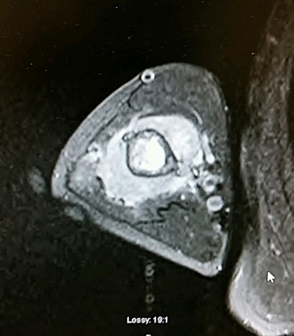
Brief description of the xray
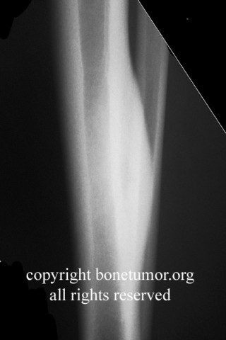
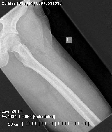
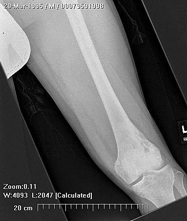
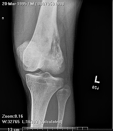
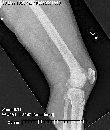
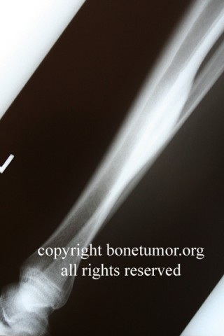
Classic radiological presentation includes radiolucent nidus surrounded by a dramatic reactive sclerosis in the cortex of the bone
Complete Information on this Tumor
Introduction and Definition
Osteoid Osteoma is a benign bone lesion with a nidus of less than 2 cm surrounded by a zone of reactive bone. This lesion accounts for approximately 10 % of benign bone tumors.
Incidence and Demographics
The tumor occurs most frequently in the second decade, with a peak age in the early twenties,and affects males twice as often as females.The proximal femur is the most common location followed by the tibia, posterior elements of the spine, and the humerus. Osteoid Osteoma is found in the diaphysis or the metaphysis of the proximal end of the bone more often than the distal end.
Symptoms and Presentation
Osteoid osteoma has a distinct clinical picture of dull pain that is worse at night and disappears within 20 to 30 minutes of treatment with non-steroidal anti-inflammatory medication. Joint pain may be present with a periarticular lesion and synovitis can occur secondary to an intraarticular lesion. Local symptoms can include an increase in skin temperature, increased sweating and tenderness. Epiphyseal lesions can cause abnormal growth.
X-Ray Appearance and Advanced Imaging Findings
The classic radiological presentation of an osteoid osteoma is a radiolucent nidus surrounded by a dramatic reactive sclerosis in the cortex of the bone. The center can range from partially mineralized to osteolytic to entirely calcified. The lesion can occur only in the cortex, in both the cortex and medulla, or only the medulla. The reactive sclerosis may be present or absent. The four diagnostic features include (1) a sharp round or oval lesion that is (2) less than 2 cm in diameter, (3) has a homogeneous dense center and (4) a 1-2 mm peripheral radiolucent zone.'
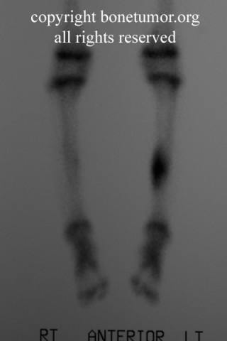
CT is the preferred method of evaluation, especially if the lesion is in the spine or obscured by reactive sclerosis. The radiologic differential includes osteoblastoma, osteomyelitis, arthritis, stress fracture and enostosis.
Histopathology findings
On gross examination, osteoid osteoma is a brownish-red, mottled and gritty lesion that is distinct from the surrounding bone. It can be present in the cortex or medullary canal. Osteoclasts are present. The nidus is surrounded by sclerotic bone with thickened trabeculae.
Microscopically, the nidus consists of a combination of osteoid and woven bone surrounded by osteoblasts. The oval shaped nidus is welvascularized and clearly separate from the reactive woven or lamellar bone.
Treatment Options for this Tumor
Osteoid osteoma will resolve without treatment in an average of 33 months. If the patient does not wish to endure the pain and prolonged use of non-steroidal anti-inflammatory medications, surgical removal or percutaneous ablation of the nucleus is indicated.
Suggested Reading and Reference
References
'Bloem, J and H. Kroon, Osseous Lesions, Radiologic Clinics of North America,31(2):261-277, March, 1993. DGitelis, S, Wilkins, R and EU
Conrad, benign Bone Tumors, Instructional Course Lectures, 45:425426, 1996.
Bullough, Peter, Orthopaedic Patholoev (third edition), Times Mirror International Publishers Limited, London, 1997.
Huvos, Andrew, Bone Tumors:Diagnosis, Treatment and Prognosis, W.B. Saunders, Co., 1991.
11/18/97