Liposclerosing Myxofibrous Tumor of bone

Summary
Description

The patient is a 44-year-old male machinist who has noticed pain in the right hip for approximately one year. This led to evaluation with imaging studies showing a lesion in the proximal femur adjacent to the lesser trochanter.

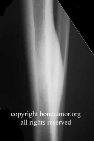
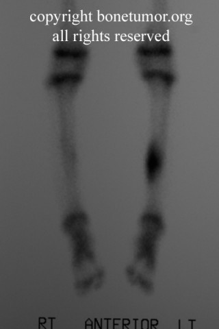
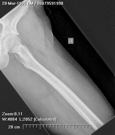
Plain radiographs, CT scan, bone scan are shown at the left. Please click on the thumbnails for a larger image. There is a mixed lytic and sclerotic lesion in the proximal femur, centrally located in the intramedullary space, just above the lesser trochanter. The cortex does not appear to be violated. The area in the center of the lesion is lucent without matrix. Surrounding this is a very dense area of sclerosis which is somewhat irregular.

Brief description of the xray
The CT scan shows essentially similar findings but shows that the lesion is larger when seen on CT that when seen on radiographs. The central area is actually two lobulated central lucent defects in the bone. The lesion is very slightly increased uptake on bone scan. No other areas of abnormality on the bone scan are appreciated.
Tumor Type
Benign or Malignant
Most Common Bones
Complete Information on this Tumor
Introduction and Definition

This tumor is usually in the proximal femur.. A diagnosis must be based on the combination of the location and appearance with the predominant histological pattern. These lesions are usually incidental findings. The age range is broad, usually adults, The tumors probably arise in childhood. Their appearance may evolve slowly over time.

Differential Diagnosis
The differential diagnosis of this lesion should include fibrous dysplasia, fibroxanthoma (non-ossifying fibroma), polymorphic fibro-osseous tumor of bone, liposclerosing myxofibrous tumor of bone, myxofibroma, lipoma, cyst, bone infarct, Paget's disease, and, chondroma. The lesion is too large for an osteoid osteoma but might be an osteoblastoma. The lesion does not have aggressive features so osteosarcoma is unlikely. No orthopedic surgeon considering a bone-forming lesion in the femur should fail to at least consider osteosarcoma, regardless of the age of the patient.
Histopathology findings
This lesion was biopsied by minimally invasive means, with an approach calculated to minimize the risk of biopsy-induced pathologic fracture as well as to minimize the risk of contamination of uninvolved structures in the case of a malignancy.

The tumor is a polymorphic fibro-osseous tumor of bone, also called a liposclerosing myxofibrous tumor of bone. On pathology, the lesion is composed of crudely woven bone that may have a pagetoid appearance. surrounded by fibrous tissue. Fat and myxoid change may also be present. The lesion may mimic fibrous dysplasia.
Treatment Options for this Tumor
The tumor may not have features that allow diagnosis without careful biopsy, thus observation only is not appropriate management of this tumor. After biosy, this tumor does not require aggressive resection. Treatment by curettage is sufficient. The patient should be followed to check for progression. In parallel with some enchondromas and bone infarcts, a minority of lesions undergo malignant transformation.
Suggested Reading and Reference
References:

Hum Pathol. 1993 May; 24(5): 505-12.

Polymorphic fibro-osseous lesions of bone: an almost site-specific diagnostic problem of the proximal femur.

Ragsdale BD.