Florid reactive periostitis

Summary
Description

Florid reactive periostitis (FRP) is a rare benign bone lesion that affects the fingers or toes of adolescents and young adults (range 5 - 70 years) Women are more commonly affected than men.

The lesion has been given a number of names, including benign fibro-osseous pseudotumor, fasciitis ossificans, parosteal fasciitis, pseudomalignant osseous tumor of soft tissue, and others. Florid reactive periostitis is the term recommended by Landsman. The fingers are equally affected. One case has been reported in the tibia.

Symptoms and Presentation
(1) The lesion appears to be a reactive process rather than a true neoplasm. The cause of FRP is unknown, but about one-half of patients with this lesion give a history of trauma to the area. The lesion is most common in the tubular bones of the hands and feet. In he hand, most lesions occur in the proximal phalanx. Lesions in the distal phalanx, the metacarpals, and the thumb are rare.

(2) Clinically , there is a history of gradually progressive swelling, erythema, and pain or a painful mass in the affected part. Approximately 50% of patients have a history of trauma. The symptoms often develop over one to two months, but there may be a longer history of mild or minimal symptoms or a long history of a nodule in the area. The symptoms are exacerbated by use or weightbearing on the affected bone, and improved by rest, antiinflammatories and corticosteroids. The mass may decrease in size if steroids are given. There are no constitutional symptoms.
Tumor Type
Benign or Malignant
Complete Information on this Tumor
Introduction and Definition

Florid reactive periostitis (FRP) is a rare benign bone lesion that affects the fingers or toes of adolescents and young adults (range 5 - 70 years) Women are more commonly affected than men.

The lesion has been given a number of names, including benign fibro-osseous pseudotumor, fasciitis ossificans, parosteal fasciitis, pseudomalignant osseous tumor of soft tissue, and others. Florid reactive periostitis is the term recommended by Landsman. The fingers are equally affected. One case has been reported in the tibia.

Symptoms and Presentation

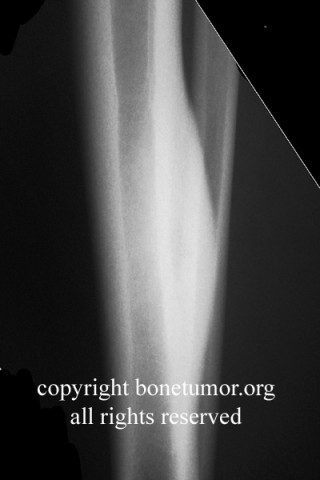
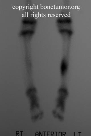
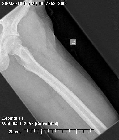
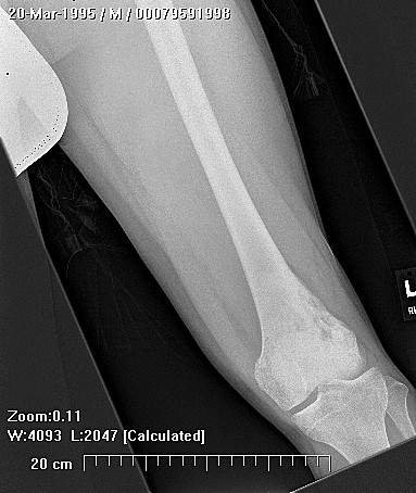
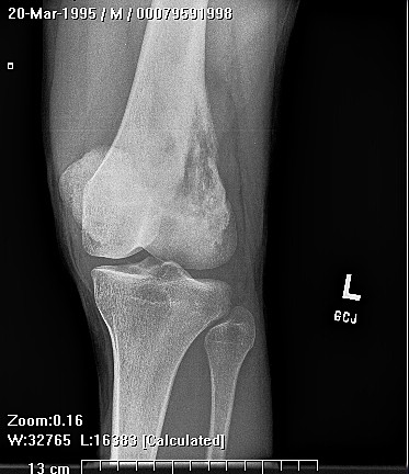
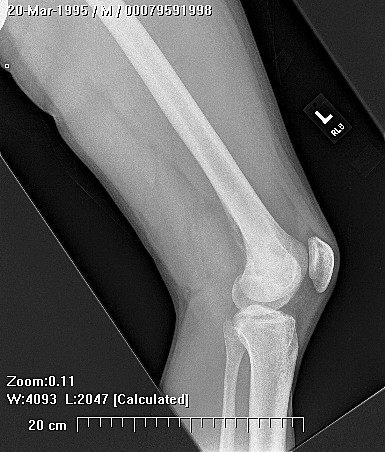
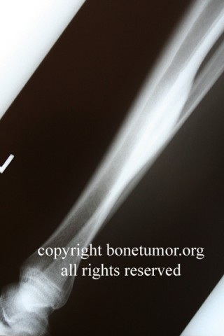
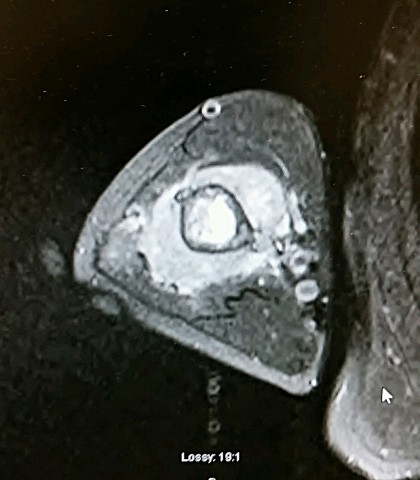
Examination reveals a tender mass with mild surrounding warmth and erythema, but no fever or adenopathy. Laboratory studies typically show a normal white blood-cell count and differential and a mildly elevated sedementation rate. Electrolytes and other laboratory values are unaffected. Plain radiographs show the tumor is adjacent to the bone rather than arising from it. Soft tissue swelling is seen as well as a marked periosteal reaction. There may be new bone formation in the soft tissues and subtle cortical thinning. In rare cases there is local erosion of the cortex. Technetium-99 bone scans show a solitary focus of intense uptake. Magnetic resonance scans show a mass and striking signal abnormalities in the soft tissues as well as the nearby bone marrow that are relatively nonspecific and are consistent with infection, trauma, or tumor.

Treatment Options for this Tumor
While the lesion is benign, the findings overlap with this of osteosarcoma, infection, and other aggressive lesions. The differential diagnosis includes conventional osteosarcoma, periosteal osteosarcoma, parosteal osteosarcoma, bizarre parosteal osteochondromatous proliferation (Nora's lesion), chondrosarcoma, giant cell tumor of tendon sheath, osteochondroma infection, stress fracture, and myositis ossificans,. Histologically, the predominant cells are large spindle shaped fibroblasts with prominent nuclei. There is no pleomorphism. Mitoses are present or even frequent but not abnormal. Mulinucleated giant cells are frequently seen. There is mature and immature osteoid and woven bone, cartilage, or a mix of chondroid, osteoid, and myxoid elements. There may be zonal maturation of the tissues as is seen in myositis ossificans. Patel and Desai () have recommended applying the Enneking staging system for benign tumors() to this lesion. Treatment consists of a careful workup followed by biopsy as well as aerobic and anaerobic cultures to rule out other potentially more worrisome lesions. Once the diagnosis has been confirmed, careful and complete surgical removal by excision through the margin of the lesion is indicated. There have been reports of recurrence following local excision, prompting recommendations of wide or radical excision as a primary treatment. However, most patient will not have a recurrence following marginal excision, thus radical surgery does not appear to warranted as a primary treatment. There have been some reports of spontaneous resolution. Recurrence after local excision does occur and may be rapid. The histological and radiographic findings do not appear to be consistently helpful in predicting which patients are more likely to have a recurrence. The likelihood of recurrence may be decreased by radiation or prostaglandin antagonists, although no data to support these treatments is available. Repeat local excision has been reported to be successful for treating recurrent lesions. In unusually aggressive or multiply recurrent cases ray amputation can be used. There have been no recurrences after delayed ray resection. To our knowledge, there have been no reports of metastasis or multifocal occurrences of this lesion. 2/15/00 HD
Suggested Reading and Reference
References: Landsman JC, Shall JF, et al: Florid Reactive Periostitis of the digits. Orthop. Rev. 1990;19:828-834 Brien EW, Zahiri CA, Mirra JM. FRP Ossificans of the Tibia. JBJS 1999;81-A:1002-1007 Howard RF, Slawski DP, Gilula LA. FRP of the Digit With Cortical Erosion. J Hand Surg. 1996;21A:501-505 Rogers GF, Brzezienski MA FRP of the Middle Phalanx. J Hand Surg. 1999;24A;1014-1018