Summary
Eosinophilic granuloma (EG) is a solitary, non-neoplastic proliferation of histiocytes. EG is part of a spectrum of Langerhan's cell histiocytosis, formerly known as histiocytosis X.
Complete Information on this Tumor
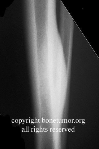
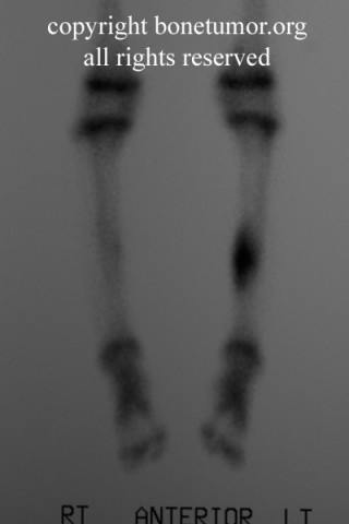
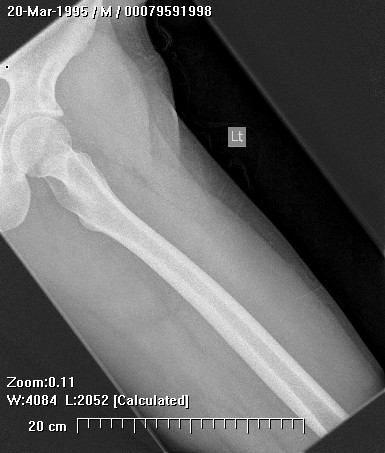
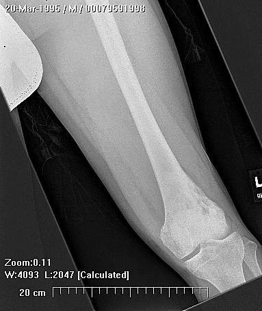
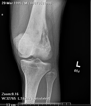
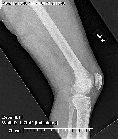
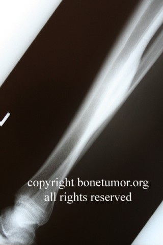
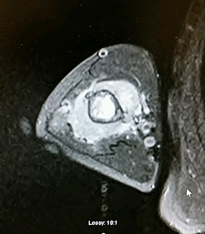
Eosinophilic granuloma (EG) is a solitary, non-neoplastic proliferation of histiocytes. EG is a localized lesion in bone or lung. EG is part of a spectrum of Langerhan's cell histiocytosis, formerly known as histiocytosis X. EG is found in the skull, mandible, spine and long bones. Letterer-Siwe disease is a fulminant systemic disease that comprises 10% of Langerhan's cell histiocytosis, occurs in children under 3 years old and is rapidly fatal. Hand-Schuller-Christian disease (HSC) is a chronic disseminated form of Langerhan' s histiocytosis and occurs in older patients. The well known triad of HSC is diabetes insipidus, exopthalmos and skull lesions. EG can convert to systemic forms of the disease. It makes up 60-80% of all cases of Langerhan's cell histiocytosis.
EG is normally symptomatic. Local pain, swelling and tenderness are common and the ESR may be elevated.
Under the microscope, EG consists of sheets of Langerhan's cells. These cells are derived from the mononuclear cell and dendritic line precursors and are found in the bone marrow. The cell is identifiable under the electron microscope as the Langerhan's cell has racket shaped cytoplasmic inclusion
bodies called Birbeck's granules. Also present in the lesion are varying amounts of lymphocytes, polymorphonuclear cells, eosinophils and giant cells. Early lesions have many Langerhan's cells and eosinophils. Older lesions have fewer cells and much fibrous tissue. The cause of EG is unknown and speculated to be either infection or immunological.
Greis, PE and FM Hankin, Eosinophilic Cranuloma, Clinical Orthopaedics and Related Research, 257:204211, August, 1990.
Conway, WF and CW Hayes, Miscellaneous Lesions of Bone, Radiologic Clinics of North America, 31(2): 339-357, March, 1993.
Bulloughs, Peter. Orthopaedic Pathologv (third edition), Times Mirror International Publishers Limited, London, 1997. Huvos, Andrew. Bone Tumors: Diagnosis. Treatment and Prognosis, W.B. Saunders, Co., 1991.