Schwannoma - neurilemmoma - foot and ankle

Summary
People and Age
Benign schwannoma/benign neurilemmoma is a solitary nerve sheath tumor that typically presents in adults between age 20 and 50
Symptoms and Presentation
Most lesions are asymptomatic. Schwannoma is rarely found in the foot. The typical solitary tumor presents as a slow growing painless mass which may have been present for 1 to 2 years or more.
Brief description of the xray
There can be local bony impingement and bone remodeling because of pressure from the tumor, but these tumors do not invade bone.
Brief desc of tx
Most lesions may be observed without surgery. Because of the indeterminate MRI appearance of this tumor, a complete evaluation and staged biopsy is recommended before definitive surgical removal is planned. Treatment is by excision with a marginal margin
Benign or Malignant
Body region
Most Common Bones
Tumor behavior
Tumor density
Complete Information on this Tumor
Introduction and Definition

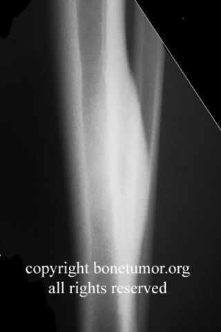
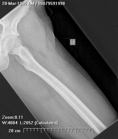
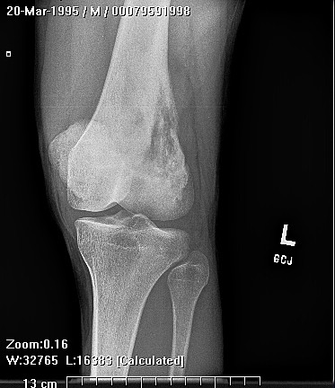
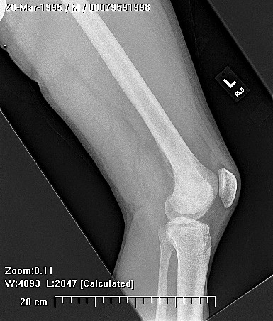
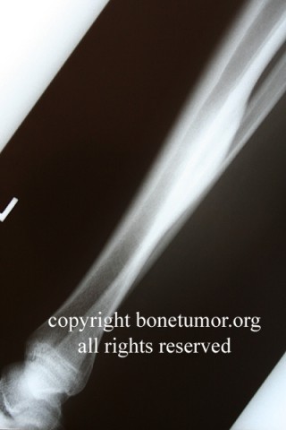
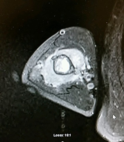
Schwannoma is also called neurilemoma, neurinoma, neurocytoma, peripheral glioma and peripheral fibroblastoma (1,2). World Health Organization Classification of Tumours (3) uses the term schwannoma, which is used here. (3). A schwannoma is a benign, encapsuled and non-invasive tumour that is derived from schwann cells (4). Schwannomas are uncommonly found in the foot (5). The etiology is unknown, but risk factors include trauma and neurofibromatosis type 2 (4). The clinical presentation depends on the location and size of the lesion. The sign and symptoms will typically result from the mass effect and / or direct involvement of the nerve and surrounding tissue (6).

Incidence and Demographics
Schwannoma affects man and women equally with peak age at diagnosis of 30–40 years, and is most commonly localized in the dermis and subcutis with a predilection for head and neck (7). In one single-center retrospective review, only 10,2% (14 of 137) were located in the foot and / or ankle (8). In another series, 12 of 104 (11,5%) during 32 years period were located in the foot and ankle (9). This well encapsulated tumor is usually benign in nature and malignant transformation is rarely reported (10).
Symptoms and Presentation

The tumors are slow growing and malignant transformation is rare(2). Most lesions are asymptomatic. The typical solitary tumor presents as a slow growing painless mass which may have been present for 1 to 2 years or more. The discovery of one schwannoma should trigger a careful search for others.

X-Ray Appearance and Advanced Imaging Findings
These tumors are not calcified and normally are not visualized by plain xrays. There can be local bony impingement and bone remodeling because of pressure from the tumor, but these tumors do not invade bone.
MRI Findings
MRI is particularly useful, it shows a usually round or oval mass with a moderately bright signal on T1-weighted images and a bright, heterogeneous signal on T2-weighted images. The mass is usually less than 2.5 cm in size. The lesion enhances uniformly with gadolinium contrast
Differential Diagnosis
synovial sarcoma
Preferred Biopsy Technique for this Tumor
incisional
Histopathology findings
The tumor has a biphasic appearance. Antoni A areas of the tumor are very cellular with a spindle shaped cell population showing nuclear palisading and scattered Verocay bodies. Antoni B areas show less cellular areas of hyalinization, hemosiderin deposition and cystic changes. Nuclear atypia of degenerative nature may be present.
Treatment Options for this Tumor
observation - excision with a marginal margin
Preferred Margin for this Tumor
marginal
Outcomes of Treatment and Prognosis
Recurrence is rare
Special and Unusual Features
May be very slow growing and presnet with an unusually long history, many years.
Suggested Reading and Reference
1. Stout AP, Carson W. The pepipheral manisfestations of the specific nerbe sheath tumours (neurilemoma). Am J Cancer 1935;24:751-89.
2. Rockwell GM, Thoma A. Schwannoma of the hand and wrist. Plast Reconstr Surg 2003;111(3):1227-32.
3. Fletcher, C. Unni, K. Mertens, F. World Health Organization Classification of Tumours. Pathology and Genetics of Tumours of Soft Tissue and Bone. IARC Press. Lyon, (2002).
4. Ferner RE, O’Doherty MJ. Neurobibroma and schwannoma. Curr Opin Neurol 2002; 15:679-84.
5. Still G. Neurilemoma of the medial plantar nerve: a case report. J Foot and Ankle Surg 2001; 40(4):236-9.
6. Mrugala M, Batchelort, Plotkin S. Peripheral and cranial nerve sheath tumors. Curr Opin Neurol 18:604-610, 2005.
7. Iwashita T, Enjoji M. Plexiform neurilemmoma: a clinicopathological and immunohistochemical analysis of 23 tumours from 20 patients. Virchows Arch A Pathol Anat Histopathol. 1987;411(4):305-9. PMID: 3114942.
8. Ruggieri M. The different forms of neurofibromatosis. Child Nerv Syst 15:295,1999.
9. Kehoe NJ, Reid RP, Semple JC. Solitary benign peripheral-nerve tumours: review of 32 years experience. J Bone Joint Surg Br 77:497-500. 1995.
10. Harkin JC, Reed RJ. Tumors of the peripheral nervous system, fascicle 3, second series. Washington, DC: Armed Forces Institute of Pathology, 1969:60-64.
11. Ghaly RF. A posterior tibial nerve neurilomoma unrecognized for ten years: case report. Neurosurgery 2001;48 (March (3)):668-72.
12. Fortnum H, O'Neill C, Taylor R, Lenthall R, Nikolopoulos T, Lightfoot G, et al. The role of magnetic resonance imaging in the identification of suspected acoustic neuroma: a systematic review of clinical and cost effectiveness and natural history. Health Technol Assess. 2009 Mar. 13(18):iii-iv, ix-xi, 1-154.