Summary
Description
Granuloma annulare (GA) is a benign inflammatory granulomatous dermatosis. GA is characterized clinically by dermal papules and annular plaques.
People and Age
GA does not favor a particular race, ethnic group, or geographical area. Localized GA is the most common among the various subtypes and occur in patients younger than 30 years.
Symptoms and Presentation
Both localized and generalized GA lesions usually manifest as asymptomatic cutaneous lesions. One or several small, firm, flesh colored or erythematous papules are seen, which may form a ring, and slowly expand in diameter.
Brief description of the xray
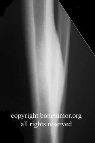
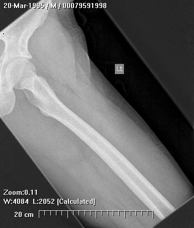
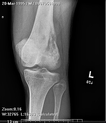
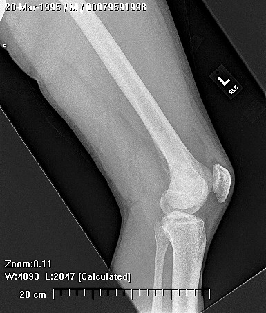
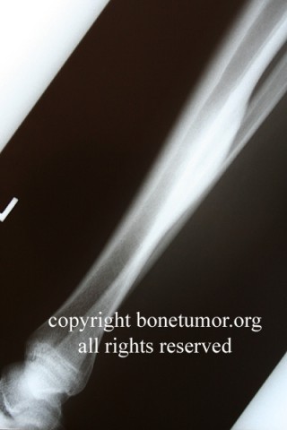
Radiographs of subcutaneous GA show a nonspecific soft tissue mass without calcification.
Brief desc of tx
No treament is needed for many cases. A wide variety of treatments have been reported but no single approach has been proven to be the best.
Complete Information on this Tumor
Introduction and Definition
Granuloma annulare (GA) is a benign, asymptomatic, inflammatory granulomatous dermatosis. The lesion usually presents as a group of plaques or papules that form an enlarging, roughly ringlike shape. GA is relatively common disease that occurs in all age groups, but it is rare in infancy. The precise cause of GA is unknown. There are many clinical variants such as localized, generalized, arcuate, subcutaneous, and perforating. GA is seen most commonly in hands and feet.
Incidence and Demographics
The frequency of GA is in the general population is unknown. GA does not favor a particular race, ethnic group, or geographical area. Localized GA is the most common among the various subtypes and occur in patients younger than 30 years . Women are affected by GA twice as often as men, subcutaneous GA is predominantly a disease of otherwise healthy children, who are typically aged 2-10 years.
Symptoms and Presentation
Localized and disseminated GA are the two most common types.
Localized GA comprises about 3 out of 4 cases. One or several small, firm, flesh colored or eyrhematous papules are seen, which may form a ring, and slowly expand in diameter. These lesions typically occur on the lateral and dorsal surfaces of the hands and feet.
Disseminated or generalized GA is characterized by widespread lesions similar in nature to those in localized GA. The lesions may last for several years. Lesions may improve in winter and worsen in summer.
Subcutaneous GA is most common in children 2 to 5 years old. This form most often manifests as a large, asymptomatic soft tissue mass. Although nodules are usually stable for months, they may rapidly enlarge over the course of weeks. Patients with subcutaneous GA present with a firm, nontender, flesh-colored or pinkish nodule without overlying epidermal alteration .Lesions are typically solitary but may occur in clusters. The most commonly reported site of involvement is the lower extremities (65% of cases), often on the pretibial surface. Other typical sites include the fingers and palms and the dorsa of the feet. The buttocks, forehead, and scalp are less commonly affected. Deep dermal or subcutaneous nodules on the extremities are attached to fascia and are often therefore mobile, whereas lesions on the scalp are attached to underlying periosteum and are therefore fixed or only slightly mobile. These lesions do not progress to a systemic illness.
Perforating is a rare type, more common in women, and found on the upper limbs and pelvis, abdomen, trunk, and extremities. The lesions may cause itching and pain.
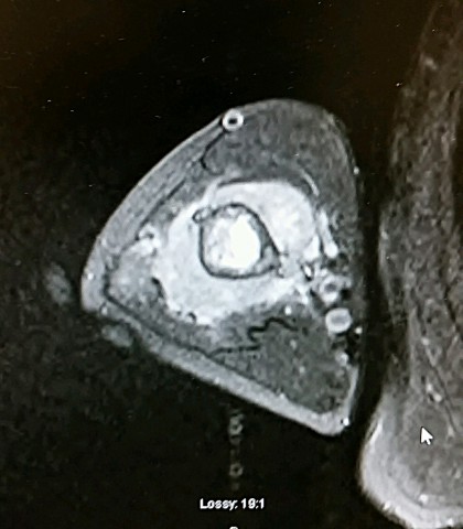
X-Ray Appearance and Advanced Imaging Findings
Radiographs of subcutaneous GA show a nonspecific soft tissue mass without calcification.On CT scans, subcutaneous GA appears as a poorly defined mass with variable attenuation and variable contrast enhancement.On sonography, GA are often ill defined and hypoechogenic with no internal flow on Doppler sonography . On MR imaging, subcutaneous granuloma annulare appears ill defined and relatively isointense relative to muscle on T1-weighted images. Signal intensity on T2- and proton density—weighted MR images can be low or heterogeneously hyperintense , These lesions may enhance after administration of contrast material.
Differential Diagnosis
The most important consideration for the foot and ankle surgeon is differentiation of GA from epitheleiod sarcoma and synovial cell sarcoma, which may have a similar presentation. For this reason biopsy of these lesions is recommended. In addition, other dermatologic disorders can mimic GA, such as Dermoid cyst, rheumatoid nodules, erythema nodosum, necrobiosis lipoidica and epithelioid sarcoma, reticulohistocytoma, reticulohistiocytic granuloma.
Preferred Biopsy Technique for this Tumor
incisional
Histopathology findings
Histopathologically, subcutaneous GA consist of areas of basophilic degeneration of collagen bundles with peripheral palisading granulomas involving the connective tissue septa of the subcutis. The central necrobiotic areas contain increased amounts of connective tissue mucin and nuclear dust from neutrophils between the degenerated collagen bundles. Eosinophils are common. Immunoflourescence studies have also shown the presence of fibrin, IgM, and C3 deposition in the lesion.
Treatment Options for this Tumor
GA is not always symptomatic and it has a tendency towards spontaneous resolution. As a result, no treatment may be needed. Painful or disfiguring lesions have been treated by various methods, although the level of evidence supporting these methods is low. Treatment include potent topical corticosteroids, as well as with intralesional corticosteroids, cryotherapy, tacrolimus and pimecrolimus and imiquimod cream, intralesional alkylating agents, and excision by surgery.
Preferred Margin for this Tumor
This tumor does not require a negative margin.
Outcomes of Treatment and Prognosis
The behavior of GA termed to be unpredictable, to date there appears to be no one therapy that has been consistently effective, local or distant recurrences have been reported in 20-75% of cases in published studies.
Special and Unusual Features
This lesion is typically asymptomatic and ultimately self-limited. In addition, it has been shown to recur in a large proportion number of cases where surgical treatment has been given. As a result, the role of surgery is limited to diagnostic biopsy procedures. Surgical treatment with curative intent appears to be unnecessary.
Suggested Reading and Reference
1-subcutaneous granuloma annulare:
Requena L, Fernández-Figueras MT.
Semin Cutan Med Surg. 2007 Jun;26(2):96-9. Review.PMID: 17544961
2-Deep granuloma annulare (pseudorhumatiod nodule) in children: clinicopathologic study of 35 cases.
McDermott MB, Lind AC, Marley EF, Dehner LP.
Pediatr Dev Pathol. 1998 Jul-Aug;1(4):300-8. Review.PMID: 10463292 .
3- Subcutaneous granuloma annulare in childhood: clinicopathologic study of 34 cases.
Grogg KL, Nascimento AG.
Pediatrics. 2001 Mar;107(3):E42.PMID: 11230623
4- Granuloma Annulare
Author: Ruby Ghadially, MBChB, FRCP(C)Derm, Professor, Department of Dermatology, University of California, San Francisco, School of Medicine
Coauthor(s): Akos Z Szabo, MD, Resident Physician, Department of Plastic and Hand Surgery, Freiburg University Medical Center; Amit Garg, MD, Director of Clinical Elective in Dermatology, Assistant Professor, Department of Internal Medicine, Division of Dermatology, University of Massachusetts Medical School
5- Sonography and MR Imaging of Selected Benign Masses in the Ankle and Foot
Hong Pham1, David P. Fessell1,2, John E. Femino3, Susan Sharp1, Jon A. Jacobson1 and Curtis W. Hayes1
1 Department of Radiology, University of Michigan Medical Center, Taubman Center (TC) 2808, 1500 E. Medical Center Dr., Ann Arbor, MI 48109-0326.
2 Present address: Akron Radiology, 525 E. Market St., Akron, OH 44304.
3 Department of Orthopedic Surgery, University of Michigan Medical Center, Taubman Center, Ann Arbor, MI 48109-0326.
6-Granuloma annulare: a case presentation of the typical and subcutaneous forms.
Miketa JP, Prigoff MM.
J Foot Ankle Surg. 1993 Jan-Feb;32(1):34-7. Review