Enchondroma - Foot and Ankle

Summary
Description

This tumor occurs in the metatarsals and phalanges.

People and Age
Occurs in the adult years, 20's - 40's
Symptoms and Presentation
Presents with pain. In the phalanges, usually after a small fracture.
Brief description of the xray
On xray the tumor is lucent with flocculent matrix calcification
Brief desc of tx
Curettage is the treatment of choice for most of these tumors.
Benign or Malignant
Body region
Most Common Bones
Location in bone
Position within the bone
Tumor behavior
Complete Information on this Tumor
Introduction and Definition

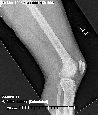
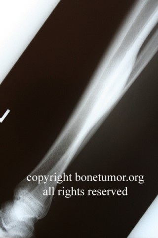
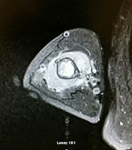
Approximately 8% of these tumors occur in the bones of the foot. Small, peripheral cartilage tumors tend to be benign, where as large central cartilage lesions are more likely to be malignant. Reliable differentiation of benign from malignant cartilage tumors is difficult. Tumors that are larger, tumors located in the hindfoot or midfoot, or new tumors presenting in a patient with a known history of enchondromatosis (Ollier's disease) have an increased risk of malignancy.

Incidence and Demographics
The peak age at diagnosisis around the middle of the fourth decade, but the tumor may present as virtually any age
Symptoms and Presentation

Patients present with pain during activities or after an injury, but there is rarely any mass palpable on physical examination. For enchondromas in the phalanges, pathological fracture through the lesion will cause the patient to seek medical care. A few patients have multiple enchondromas and warrant special attention and possibly referral to a bone tumor specialist.

X-Ray Appearance and Advanced Imaging Findings
Enchondroma usually occurs in the metatarsals or phalanges of the lesser toes. The hindfoot is rarely involved. The lesion is typically a solitary, slightly expansile,lucent lesion with minimal matrix calcification, that may thin, expand, or fracture the nearby cortex. Radiographically, the lesion is expansile and lytic, with a variable amount of matrix mineralization. The matrix has been described as "popcorn" or "stippled". The matrix consists of nodules of cartilage which tend to calcify at the periphery, so that on high quality radiographs "ring and arc" forms are visible. Clear demonstration of these helps confirm the diagnosis so that biopsy is not necessary. Other radiological findings such as the presence or absence of matrix mineralization, amount and extent of cortical thinning or expansion, and findings on technetium bone scans do not seem to be well correlated with the presence or absence of malignancy.
Differential Diagnosis
Juxtacortical chondroma, chondromyxoid fibroma. Giant cell tumor may be mistaken for enchondroma, and both can occur in the foot in similar locations. GCT has no matrix calcification and occurs exclusively in the metaphysis and epiphysis adjacent to the growth plate, often extending right up to the joint surface. In small bones that are completely filled with tumor, these lesions may be difficult e to differentiate. The MRI signal patterns are distinct. Treatment is very different so the surgeon should be careful to make an accurate diagnosis.
Preferred Biopsy Technique for this Tumor
Open, combined with excision after frozen section
Treatment Options for this Tumor
Treatment depends on how expansile the lesion has become. Lesions inside the medullary cavity may remain totally latent, are usually asymptomatic, and no treatment other than intermittent radiographic follow-up is indicated. If a medullary enchondroma requires biopsy for some reason, it can be curetted at the same time and the defect filled with an apropriate bone void filler. Extremely expansile lesions may require complete excision and substitution of a structural allograft. In the foot, these lesions can cause cosmetic and functional loss and pathological fracture. Large lesions in the distal phalanges should be considered for partial amputation of the toe, since the functional and cosmetic result of curettage and bone grafting may be unacceptable.
Preferred Margin for this Tumor
Intralesional
Outcomes of Treatment and Prognosis
Enchondromas recur very rarely following treatment, so that total eradication of the lesion is not necessary, rather the goal should be is restoring appearance and function. Curettage and bone grafting is usually adequate.
Special and Unusual Features
Enchondroma may be highly expansile, especially in small bones such as the phalanges, so that the bone may be completely involved, deformed, and even destroyed. This is not considered a sign of malignancy in enchondromas of the foot or hand. This same behavior seen in a long bone such as the femur or humerus would be interpreted as a strong suggestion of malignancy.