Mesenchymal chondrosarcoma

Summary
Description

This rare form of chondrosarcoma is a deadly high grade sarcoma consisting of primitive mesenchymal cells and islands of cartilage which occurs most often in the femur, ribs, jaw, skull, and spine, and may occur in any bone.

People and Age
Most patients are under 40, and may be from 10 to 70. Males and females are equally affected.
Symptoms and Presentation
Patients present with pain and a mass.
Brief description of the xray
The lesions often have stippled density characteristic of cartilage tumors. They may be expansile and locally destroy bone.
Brief desc of tx
Complete wide resection, with aggressively wide margins. Long-term follow up is needed. Chemotherapy and radiation are used when complete resection is not possible, but the role of chemotherapy is not well defined. Multi-modal treatment may be considered in some centers. Chemotherapy appears to be linked to better survival.
Tumor Type
Benign or Malignant
Most Common Bones
Tumor behavior
Tumor density
Complete Information on this Tumor
Introduction and Definition

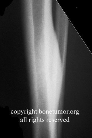
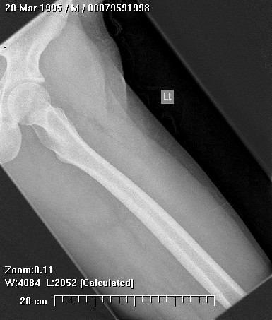
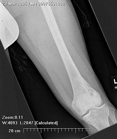
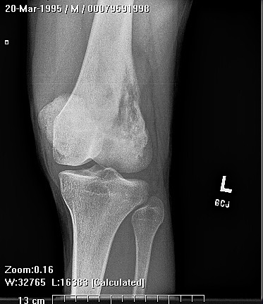
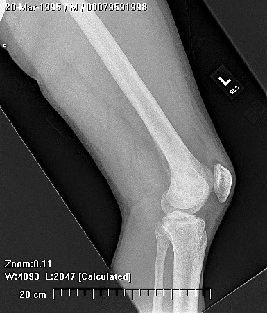
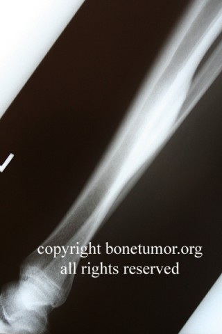
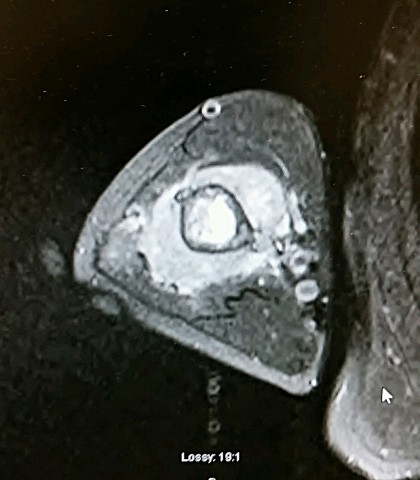
This rare form of chondrosarcoma is a tumor of undifferentiated mesenchymal cells mixed with areas of cartilage. It occurs most often in the femur, ribs, jaw, skull, and spine, and may occur in any bone. This form of chondrosarcoma is usually high grade, and the tumor is characterized by more frequent local recurrence and metastasis and a relatively poor prognosis. The xray features are not diagnostic but the histopathology is constant.

Incidence and Demographics
This tumor is very rare, accounting for about 2 - 10% of chondrosarcomas. Most patients are under 40, most are 10 to 40, and may be from 5 to 75. Males and females are equally affected.
Symptoms and Presentation

Patients present with local pain and a mass. Headache occurs in the skull lesions. Base of skull lesions present with brainstem symptoms, and cranial nerve dysfunction.

X-Ray Appearance and Advanced Imaging Findings
The lesions are predominately lytic and often have stippled density characteristic of cartilage tumors. The border is poorly defined, but may be sharp with sclerosis. They may be expansile and locally destroy bone. A significant, lobulated extraosseous mass often develops over time. Size may vary from 1 to 40 cms. This lesion may occur in the soft tissues, such as skin, joint tissues, viscera, or meninges.
Differential Diagnosis
Conventional chondrosarcoma, osteosarcoma, chondroblastoma,
Preferred Biopsy Technique for this Tumor
According to location and extent.
Histopathology findings
Sheets of spindle cells, and/or ovoid/round cells which are markedly uniform in size. Pleomorphism is very modest, with few mitotic figures. Multifocal areas of differentiation into cartilage. The undifferentiated cells predominate, the cartilage areas may make up 10 to 50% of the tumor. (Mirra) The cartilage areas stain blue, whereas they are typically pink in CBMA or CMF. The degree of anaplasia in the cartilage is variable, but usually low. Some tumors may be quite vascular, and some areas may have anastomosing vessels and resemble hemangiopericytoma, but hemangiopericytoma does not produce cartilage. Look for islands of cartilage in sheets of spindle cells or ovoid cells
Outcomes of Treatment and Prognosis
Prognosis is poor compared to most chondrosarcomas. 10 year survival is cited as 30%. Tumors tend to be high grade. Late metastasis may occur (>10 years), metastatic sites include lung, bone, viscera, and lymph nodes. Radical or very aggressive surgical treatment has been recommended. Chemotherapy as given for Ewing sarcoma has been recommended. In one of the largest series, Cesari, Mercuri et al from the Rizzoli institute found that if complete surgical remission was achieved, survival was 27%, and if it was not, survival was 0%. Chemotherapy and complete surgical remission used together were associated with a 10 year DFS of 72%, compared to 17% for surgery alone.
Special and Unusual Features
Location in non-long bone sites is more common in this tumor.
Suggested Reading and Reference
Mirra, Bone Tumors, 1989 pp577-589, AFIP fascicles, Atlas of Tumor Pathology Fechner and Mills, 1993. Cesari M, Bertoni F, Bacchini P, Mercuri M, Palmerini E, Ferrari S Tumori. 2007 Sep-Oct;93(5):423-7.
Mesenchymal chondrosarcoma. An analysis of patients treated at a single institution.