Case Identification
Case ID Number
Tumor Type
Body region
Position within the bone
Periosteal reaction
Benign or Malignant
Clinical case information
Case presentation
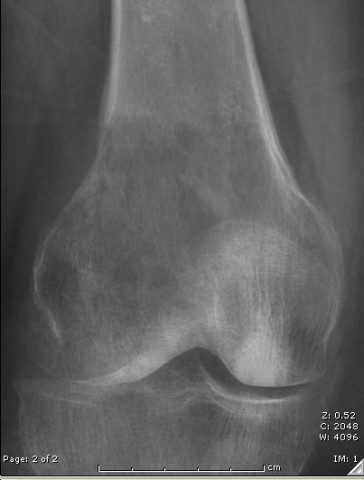
The patient is 63. She has pain in the right knee which has been increasing gradually for the past 4 or 5 months. It is now very intense. Initial x-rays (shown) were unimpressive. Later a lesion in the right distal femur was discovered. Because of the patient's medical history a large number of diagnostic possibilities exist.
Radiological findings:
Radiological findings include a large somewhat permeative mass in the right distal femur. Review of the x-rays from February and from March revealed that they were basically normal. The MRI shows a large lesion that extends into the soft tissues in the posterior portion of the distal right femoral metaphysis. There is a modest periosteal reaction. A CT scan of the chest does not show any definite abnormality. The patient sustained a pathological fracture through the lesion and was admitted to the hospital on an emergent basis. A biopsy has been performed and results are pending
Laboratory results:
No significant abnormality.
Differential Diagnosis
The patient's medical history reveals several significant conditions. She is a long-term smoker, she has a history of a pituitary mass, which was treated with radiation and completely resolved. The prompt resolution after radiation lead her doctors to presume that the lesion was a lymphoma but this was not confirmed pathologically.
Following the radiation for the pituitary mass she had panhypopituitarism requiring replacement hormones.
She has a history of a meningioma. She has a history of high-dose steroids and this led to multi-site avascular necrosis of bone, including left proximal humerus and both femoral heads, which required bilateral total hip replacements.
Finally, she has a history of a mass in the right neck which after biopsy was found to be a low grade follicular lymphoma. Her oncologist has stated that no treatment was necessary for this particular lesion
Further Work Up Needed:
After the biopsy, the challenge will be to reconstruct this badly damaged bone segment in a way that is durable, with rapid return to function and the fewest possible complications. can the bone be saved and repaired or should it be replaced?
Pathology results:
The sections reveal a monotonous infiltrate of mixed small and large lymphocytes with associated crush artifact and area of extensive necrosis. Immunohistochemical stains reveal: Positive staining for: LCA (CD45) CD20 CD10 Bcl-2 CD15, scattered positivity (hampered by necrosis and associated acute inflammation) Ki-67, approximately 50% Negative staining for: cytokeratin AE1/3, CD3, CD5, cyclin D1, CD30. There are at least 10 large cells/high power field.
Treatment Options:
After the biopsy, the challenge will be to reconstruct this badly damaged bone segment in a way that is durable, with rapid return to function and the fewest possible complications. can the bone be saved and repaired or should it be replaced?
Special Features of this Case:
An unusually complicated number of neoplastic and metabolic conditions and cancer risk factors are found in this patient, and the clinician is challenged to make a differential that includes a broad range of entities.
Image
Secret Tumor Name
Case ID Number
Image Types
Image modality
Tumor Name
Example Image
yes
Tumor Type
Benign or Malignant
Body region
Bone name
Location in the bone
periosteal reaction
position within the bone
Tumor behavior
Tumor density


