Case Identification
Case ID Number
Tumor Type
Body region
Position within the bone
Periosteal reaction
Benign or Malignant
Clinical case information
Case presentation
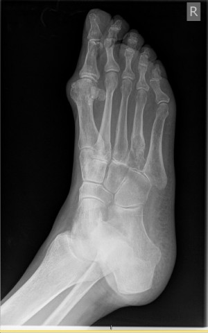
The patient a delightful 82-year-old woman, who is seen after referral from Dr. P, after a bone lesion in the base of the fourth metatarsal was found.
Radiological findings:
The patient has no personal history of malignancy. She has had pain with activity for approximately 4 months. She states that she has no pain at rest. The pain has gotten a little bit more severe. A lytic destructive and slightly expansile bone lesion has been discovered at the locus of the pain, and the base of the fourth metatarsal.
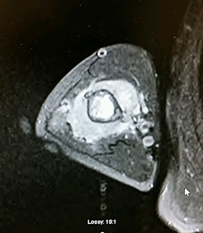
Plain radiographs show a slightly expansile, lucent, lytic lesion, with a mildly well-defined border, no sclerosis. It occupies the metaphyseal proximal portion of the fourth metatarsal. There is no extraosseous extension. The cortex is focally thinned, expanded and absent. No definite matrix mineralization is seen. Along the lateral border of the fourth, a fairly mature irregular periosteal thickening is seen, but I am not certain that this is a reaction to the tumor.
The CT scan shows a lytic expansile lesion.
A bone scan shows that there is degenerative changes in the lumbar spine no other definite abnormalities. The bone lesion in the right fourth metatarsal shows increased uptake on bone scan.
Laboratory results:
"M" SPIKE (IGM/KAPPA) = 0.48 G/DL
COMPARED TO 0.61 G/DL ON 11/15/07.
COMPARED TO 0.61 G/DL ON 11/15/07.
Differential Diagnosis
Diagnostic possibilities include both benign and malignant entities. A cartilage lesion, such as enchondroma, low grade chondrosarcoma, clear-cell chondrosarcoma might appear in this fashion. A malignancy such as multiple myeloma would be very unlikely in this location but is a possibility. The patient apparently has some history of a monoclonal spike. A metastatic malignancy with a primary location the foot in no other obvious abnormality is a possibility. Primary tumors in the kidney, gastrointestinal and genitourinary tract, lung, have been known to metastasize to the foot. Metastasis to the foot with an unknown primary is an exceedingly rare entity.
Further Work Up Needed:
Biopsy appears to be the best next step.
Pathology results:
pending
Treatment Options:
depends on pathological findings
Special Features of this Case:
In bone lesions, I recommend open biopsy over core needle and fine-needle techniques. I believe that the likelihood of having a definitive diagnosis after one procedure is significantly higher. In addition, the orthopedic oncologist can control all aspects of the biopsy process to ensure that complications are minimized. Realistically, open biopsy can be much more rapidly scheduled and completed then any type of biopsy done by a secondary service such as radiology, because of the length of time required to coordinate the intervention with a secondary service. Scheduling of the simultaneous availability of a CT scanner or floro unit, radiology technician, radiologist, and pathologist can take dozens of telephone calls, and weeks of time. All these resources are routinely available in the operating room and can be deployed within hours, if needed. If dramatic strides were made in the ease of scheduling and coordination of these procedures, they could be done as a core needle technique with acceptable diagnostic yield and probable lower cost. For the present, biopsy in the operating room is the best choice.
Image
Case ID Number
Image Types
Image modality
Tumor Name
Tumor Type
Benign or Malignant
Body region
Bone name
Location in the bone
periosteal reaction
position within the bone
Tumor behavior
Tumor density