A lesion in the talus of an 88 year old woman with 2 previous tumors Secret Tumor Name Non Hodgkin lymphoma Case ID Number 20110208LT Image Types Auxiliary Image Image modality path image Tumor Name Lymphoma of bone - foot and ankle Example Image yes Tumor Type unknown tumor - small round blue cells Benign or Malignant Malignant Body region ankle and foot, leg
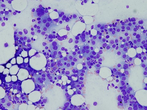
A woman in her 50's with nausea, fatigue, lethargy, and a right femoral bone lesion Secret Tumor Name Multiple myeloma Case ID Number 20100512NF Image Types Main Image Image modality path image Tumor Name unknown tumor Tumor Type unknown tumor - small round blue cells Benign or Malignant Malignant Body region knee, thigh, distal femur, proximal tibia Bone name femur Location in the bone metaphyseal periosteal reaction present position within the bone central Tumor behavior aggressive Tumor density lytic
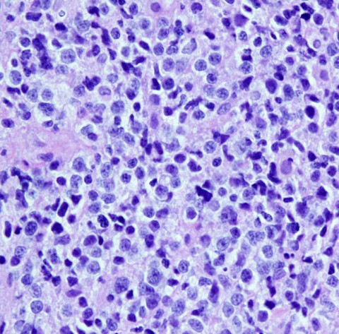
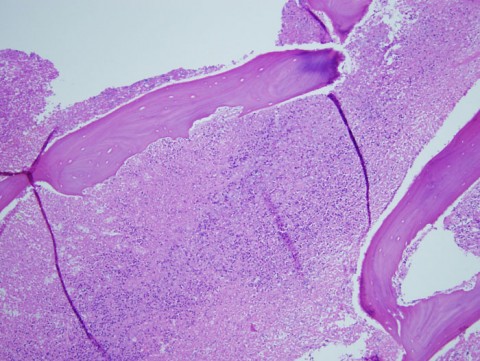
H&E 40x showing permeation of the bone by a monotonous "small blue cell tumor" associated with necrosis Secret Tumor Name Lymphoma of bone Case ID Number 20100506IP Image Types Auxiliary Image Image modality path image Tumor Name Lymphoma of bone Tumor Type unknown tumor - small round blue cells Benign or Malignant Malignant Bone name femur
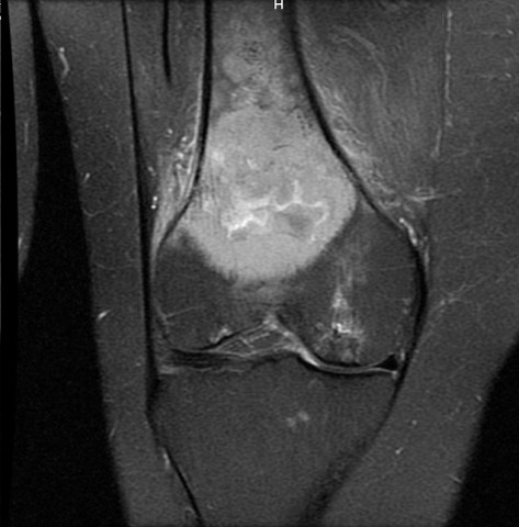
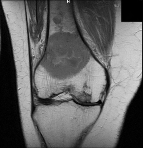
Intense pain in the knee and a permeative lesion - additional studies Secret Tumor Name Lymphoma of bone Case ID Number 20100506IP Image Types Auxiliary Image Image modality MRI Tumor Name unknown tumor Tumor Type unknown tumor - small round blue cells Benign or Malignant unknown Body region knee, thigh, distal femur, proximal tibia Bone name femur Location in the bone metaphyseal
Intense pain in the knee and a permeative lesion - additional studies Secret Tumor Name Lymphoma of bone Case ID Number 20100506IP Image Types Auxiliary Image Image modality MRI Tumor Name unknown tumor Tumor Type unknown tumor - small round blue cells Benign or Malignant unknown Body region knee, thigh, distal femur, proximal tibia Bone name femur Location in the bone metaphyseal
Intense pain in the knee and a permeative lesion - additional studies Secret Tumor Name Lymphoma of bone Case ID Number 20100506IP Image Types Auxiliary Image Image modality MRI Tumor Name unknown tumor Tumor Type unknown tumor - small round blue cells Benign or Malignant unknown Body region knee, thigh, distal femur, proximal tibia Bone name femur Location in the bone metaphyseal
Intense pain in the knee and a permeative lesion - additional studies Secret Tumor Name Lymphoma of bone Case ID Number 20100506IP Image Types Auxiliary Image Image modality MRI Tumor Name unknown tumor Tumor Type unknown tumor - small round blue cells Benign or Malignant unknown Body region knee, thigh, distal femur, proximal tibia Bone name femur Location in the bone metaphyseal
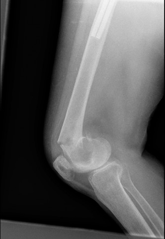
Intense pain in the knee and a permeative lesion - pathological fracture Secret Tumor Name Lymphoma of bone Case ID Number 20100506IP Image Types Auxiliary Image Image modality Xray Tumor Name unknown tumor Tumor Type unknown tumor - small round blue cells Benign or Malignant unknown Body region knee, thigh, distal femur, proximal tibia Bone name femur Location in the bone metaphyseal
Intense pain in the knee and a permeative lesion - pathological fracture Secret Tumor Name Lymphoma of bone Case ID Number 20100506IP Image Types Auxiliary Image Image modality Xray Tumor Name unknown tumor Tumor Type unknown tumor - small round blue cells Benign or Malignant unknown Body region knee, thigh, distal femur, proximal tibia Bone name femur Location in the bone metaphyseal
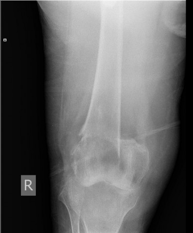
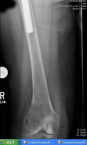
Intense pain in the knee and a permeative lesion - third xray series Secret Tumor Name Lymphoma of bone Case ID Number 20100506IP Image Types Auxiliary Image Image modality Xray Tumor Name unknown tumor Tumor Type unknown tumor - small round blue cells Benign or Malignant unknown Body region knee, thigh, distal femur, proximal tibia Bone name femur Location in the bone metaphyseal periosteal reaction present position within the bone central Tumor behavior aggressive Tumor density lytic