Case Identification
Case ID Number
Tumor Type
Body region
Position within the bone
Benign or Malignant
Clinical case information
Case presentation
The patient is a generally active 73-year-old gentleman who is retired. He has prostate cancer that has been well controlled. Now, he has right hip pain and lesions in the acetabulum and proximal femur.
Radiological findings:
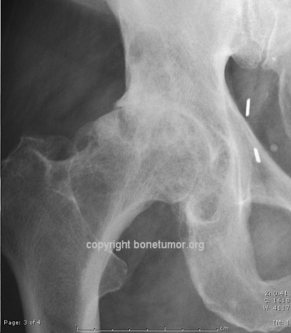
On both sides of the right hip, along with joint space narrowing and bone-on-bone contact, there are well defined cystic lesions, with sclerotic rims, which abut the joint on both sides. MRI scan the scan shows extensive abnormality in the right hip and acetabulum and supraacetabular region as well as the right femoral head. There was a cystic focus, joint space narrowing, and perilesional edema. The cystic lesions have a well defined dark rim.
Laboratory results:
The patient's prostate cancer is currently staged T3, N0, with suspicion of metastatic disease. PSA 19 at the time of treatment. Current PSA is 1.3.
Differential Diagnosis
Subchondral cyst versus metastasis or new primary malignancy.
Treatment Options:
Please see our page on subchondral cyst.
Special Features of this Case:
To differentiate between subchondral cyst and a true bone tumor or a metastatic cancer deposit in bone, look for the following features: 1) The lesion is right next to the joint. Careful examination of the radiographs may reveal an actual communication between the joint space and the cyst cavity. If doubt about the nature of the lesion exists, a fine cut CT scan on the area may allow this communicating opening to be seen and help establish the true diagnosis. 2) There are radiographically visible signs of osteoarthritis, usually moderate but sometimes mild, seen in the adjacent joint. If these are entirely absent, the diagnosis should be reconsidered. In the hip, these lesions occur the acetabulum in women with "shallow hips" which can be determined by calculating the center-edge angle. The shallow hip is prone to early degenerative changes and cysts are common in these patients. 3)There is usually a sclerotic rim around some areas of the lesion. The zone of transition is narrow, whereas in a metastatic lesion a sclerotic rim is absent and the zone of transition may be poorly defined. 4) The lesion should be fluid filled, and this may be seen best on MRI images. 5) These lesions are rarely progressive, and pathological fractures are rare.
Image
Case ID Number
Image Types
Image modality
Tumor Name
Example Image
yes
Tumor Type
Benign or Malignant
Body region
Location in the bone
periosteal reaction
position within the bone
Tumor behavior
Tumor density


