Case Identification
Case ID Number
Tumor Type
Body region
Position within the bone
Benign or Malignant
Clinical case information
Case presentation
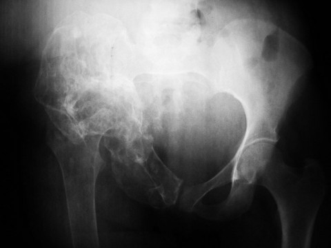
This is a consult from an ortho clinic in Kenya, a 62 y/o F presents with a transverse, midshaft femur fracture from a low-energy trauma (mechanical fall) about one week ago. She has a history of chronic hip pain for several years. She had been ambulatory prior to the fall. Additionally, she has a very bizarre-appearing hemipelvis and hip joint ipsilaterally.
Radiological findings:
The hemipelvis is almost entirely involved, diffusely enlarged, with the cortex thinned, scalloped, and there appears to be old fractures of the pubic rami. The hemipelvis is normally aligned with the normal left side, and the overall size of the right pelvis is similar to the left, suggesting a process that started after skeletal maturity, or did not interfere with the initial growth development of the hip/pelvis and proximal femur.
The Acetabulum is enlarged and thinned, but can still be seen, and is approximately spherical. The acetabulum is proximally located in the pelvis, as if a gradual process of erosion, expansion, protrusion and migration of the center of rotation occurred over a period of years. The process reaches fully to the symphysis pubis, but does not cross it, instead leaving the nearby left pubic symphysis entirely normal. The tumor is best seen in the area of the pubic symphysis on the right, with cortical thinning, expansion, areas of condensation of bone or mineral, and other areas where there is lucency. No periosteal reaction or extraosseous mass is seen, and neither is there any area where the bone is completely destroyed.
On the femoral side, the joint is gone, and an old fracture of the proximal femur/neck is seen, which has resulted in fragmentation and extrusion of the superior portion of the neck and most of the head. However, the proximal femur, up to the level of the greater troch/base of the neck, looks pretty normal in shape and development except for the osteopenia.
Laboratory results:
Her Hct is 39 and chemistries are normal (calcium
hasn’t been done). Her INR is 1.5, which is a little surprising since
she isn’t on coumadin. CXR was clean with no mets. She has no palpable
soft tissue mass in the area.
hasn’t been done). Her INR is 1.5, which is a little surprising since
she isn’t on coumadin. CXR was clean with no mets. She has no palpable
soft tissue mass in the area.
Differential Diagnosis
Please submit a three-item ddx. Explain your reasoning for each listed diagnosis. See below for email address.
Further Work Up Needed:
Biopsy of the pelvic lesion, at some superficial location. A retrograde rod is an excellent choice for the femur.
Pathology results:
Pending an update from Kenya.
Treatment Options:
Given a primary bone lesion, the bone fragility would eventually lead to protrusio of the head, (think shepards crook) then eventually a fracture, say 10 or more years ago, and with many years of walking on a non-united femoral neck fracture, this appearance could be the late result. Most likely, there nothing of interest in the femur at the site of the fracture, a biopsy there is not recommended.
Special Features of this Case:
We invite your input on this case. Send 3-item ddx to - questions (at :P) bonetumor (dot :P) org. Leave out the smileys!
Image
Secret Tumor Name
Case ID Number
Image Types
Image modality
Tumor Name
Example Image
yes
Tumor Type
Benign or Malignant


