shoulder, humerus, upper arm
Case ID Number
Image Types
Image modality
Tumor Name
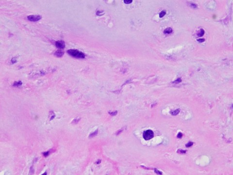
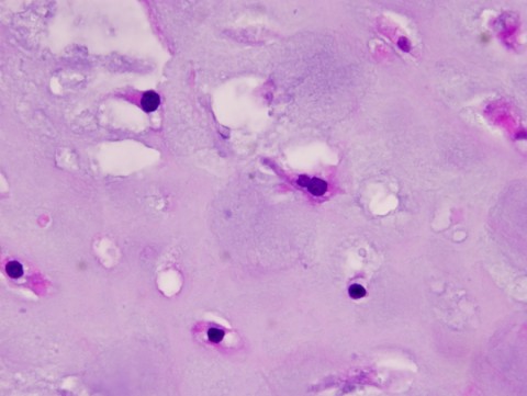
Example Image
yes
Tumor Type
Benign or Malignant
Body region
Bone name
periosteal reaction
position within the bone
Tumor behavior
Tumor density

Case ID Number
Image Types
Image modality
Tumor Name
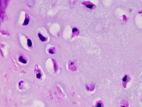
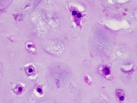
Example Image
yes
Tumor Type
Benign or Malignant
Body region
Bone name
periosteal reaction
position within the bone
Tumor behavior
Tumor density
Case ID Number
Image Types
Image modality
Tumor Name
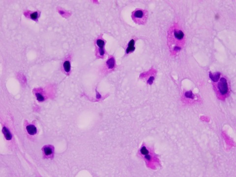
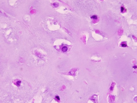
Example Image
yes
Tumor Type
Benign or Malignant
Body region
Bone name
periosteal reaction
position within the bone
Tumor behavior
Tumor density
Case ID Number
Image Types
Image modality
Tumor Name
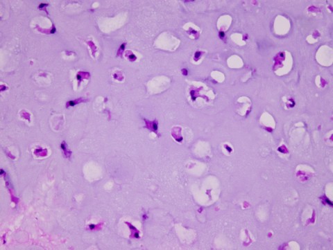
Example Image
yes
Tumor Type
Benign or Malignant
Body region
Bone name
periosteal reaction
position within the bone
Tumor behavior
Tumor density
Case ID Number
Image Types
Image modality
Tumor Name
Example Image
yes
Tumor Type
Benign or Malignant
Body region
Bone name
periosteal reaction
position within the bone
Tumor behavior
Tumor density
Case ID Number
Image Types
Image modality
Tumor Name
Example Image
yes
Benign or Malignant
Body region
Bone name
periosteal reaction
Tumor behavior
Tumor density
Case ID Number
Image Types
Image modality
Tumor Name
Example Image
yes
Tumor Type
Benign or Malignant
Body region
Bone name
periosteal reaction
position within the bone
Tumor behavior
Tumor density
Case ID Number
Image Types
Image modality
Tumor Name
Example Image
yes
Tumor Type
Benign or Malignant
Body region
Bone name
periosteal reaction
position within the bone
Tumor behavior
Tumor density


