Case Identification
Case ID Number
Tumor Type
Body region
Position within the bone
Periosteal reaction
Benign or Malignant
Clinical case information
Case presentation
A 21 year old student has had pain in the left heel for two years and is otherwise healthy. Examination of the left foot shows slight swelling in the medial and lateral border of the calcaneus. There is point tenderness over the Achilles insertion and the posterior calcaneus.
Radiological findings:
There is no discoloration of skin and no cafe au lait spots or other skin stigmata.
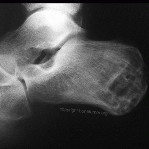
Plain x-rays, and MRI of the calcaneus are examined. There is a lesion in the posterior calcaneus which fills the bone from medial to lateral and almost fills the bone from proximal to distal. It appears to have originated in the calcaneus adjacent to the posterior Growth Plate. There are multiple lytic partial confluent lesions within bone with no defined sclerosis surrounding these lytic areas. There is no matrix mineralization. No periosteal reaction is seen.
The MRI shows the lesion in the same location which is bright on T-2 sequences and dark on T1 sequences. There are definite fluid fluid levels and multiple loculated cystic appearing lesions that are confluent. The lesion extends to the cortical border of the bone and appears to be in the cortex in some areas. No definite breakthrough or soft tissue extension is seen. No other bony abnormality is seen in the other bones of the foot.
Differential Diagnosis
This lesion appears to be a benign active or low-grade malignant bone tumor. Possibilities include Chondromyxoid Fibroma, Aneurysmal Bone Cyst, Desmoplastic Fibroma, Osteoblastoma, Chondroblastoma. A malignancy cannot be ruled out. The lesion does not appear to meet criteria for a possible Chondrosarcoma, Synovial Sarcoma, or osteosarcoma.
Image
Case ID Number
Image Types
Image modality
Tumor Name
Tumor Type
Benign or Malignant
Body region
Bone name
Location in the bone
periosteal reaction
position within the bone
Tumor behavior
Tumor density


