Case Identification
Case ID Number
Tumor Type
Body region
Benign or Malignant
Clinical case information
Case presentation
A generally healthy 19-year-old woman presents with an 11 month history of a painless ankle mass.
Radiological findings:
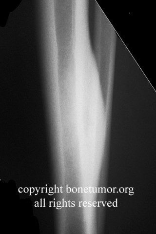
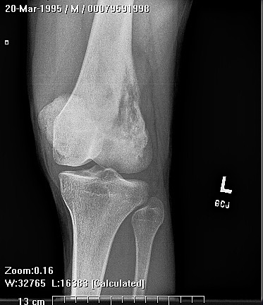
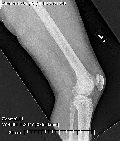
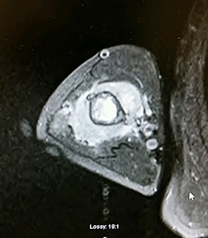
The x-ray is normal other than a soft tissue density posterior to the ankle at the level of the talus. The mass has density similar to that of fact, no calcifications. The appearance of the ankle joint and subtalar joint are normal. The MRI shows a heterogeneous mass posterior to the subtalar joint and ankle joint at the level of the talus, which measures 60 x 70 x 20 mm. It has low signal intensity on both T1 and T2 weighted sequences. No gradient echo sequences were performed. There appears to be some magnetic susceptibility artifact in some of the sequences, but this is not commented on by the radiologist. The mass is adjacent to the flexor digitorum longus, peroneal tendons, and neurovascular bundle. It lies on the surface of the posterior talus and extends from it, but there is no definite bony damage.
Laboratory results:
No laboratory examinations are made.
Differential Diagnosis
A soft tissue mass adjacent to the bones of the ankle without calcification. Possibilities include sarcoma, benign soft tissue neoplasm, a reactive process such as a synovial mass or PVNS, and other.
Further Work Up Needed:
Biopsy is necessary.
Pathology results:
biopsy is pending.
Imagen

Case ID Number
Image Types
Image modality
Tumor Name
Example Image
yes
Tumor Type
Benign or Malignant
Body region