Case Identification
Case ID Number
Tumor Type
Body region
Benign or Malignant
Clinical case information
Case presentation
A 28 year old man from northern Italy complained of mild difficulty walking for one year. He did not complain of pain. The patient had no history of unusual travel or exposure.
Radiological findings:
The vital signs were normal. No fevers had been recorded. On examination, the patient appeared comfortable and moved easily without obvious pain. There was no gross defect in the patient's ability to ambulate. There was no clonus. However, an examining neurologist had noted normal sensorium, left sided lower extremity hyperreflexia, and bilateral abnormal Babinski reflexes.
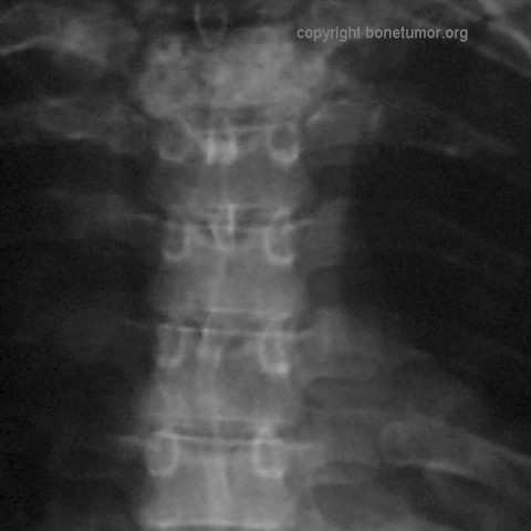
A radiograph of the chest revealed multiple pulmonary nodules, and a lesion in the second thoracic vertebrae.
Further radiographic examination of the spine showed that the lesion in T2 had caused extensive anterior and posterior changes and a soft tissue abnormality adjacent to the right side of the vertebral body. A plain radiograph, CT scan of T2, and a saggital MR image of the spinal column are shown. A CT images of the lungs are shown. A bone scan showed abnormal uptake in the region of T1 - T3 in the spine but no other abnormality (not shown).
Laboratory results:
The WBC was 10.70 (4.5 - 9.5) RBC was 5.12 (4.5 - 5.9) the hematocrit was 15.3 (14 - 18) platelets were 249 (130 - 400). There were 82.9% neutrophils, 10.9% lymphocytes, and 5.94% monocytes. Electrolytes, liver enzymes, alkaline phospate, and PT/PTT were normal. ESR was 6 (normal, less than 15) and C-reactive protein was negative (less than 0.5). There were no Bence-Jones proteins in the serum or urine. Urinalysis was normal. TSH was 0.78 (0.30 - 4.00) and TT4 was 12.4 (8.0 - 20.0). The patient was negative for HIV. A Mantoux skin test was non-reactive (normal). VDRL and TPHA were negative (no evidence for siphylis).
Pathology results:
An open biopsy of the spinal lesion was performed. Grossly, there was no purulence or granulomatous changes nor any soft tissue mass or obvious tumor. There was discoloration of the bone making it appear dark grey or black, similar to the black discoloration from metal debris from a failed orthopedic implant.
Microscopically, the lesion contained irregular anastomosing vascular channels. The channels are lined by plump endothelial cells without pleomorphism or mitotic activity. Both the background stroma and the cells lining the vascular channels stain positive for reticulum.
Microscopically, the lesion contained irregular anastomosing vascular channels. The channels are lined by plump endothelial cells without pleomorphism or mitotic activity. Both the background stroma and the cells lining the vascular channels stain positive for reticulum.
Special Features of this Case:
What is the diagnosis?
What treatment would you propose?
What do the lung nodules represent?
What treatment would you propose?
What do the lung nodules represent?
Image
Case ID Number
Image Types
Image modality
Tumor Name
Tumor Type
Benign or Malignant
Body region
Bone name


