Case Identification
Case ID Number
Tumor Type
Body region
Benign or Malignant
Clinical case information
Case presentation
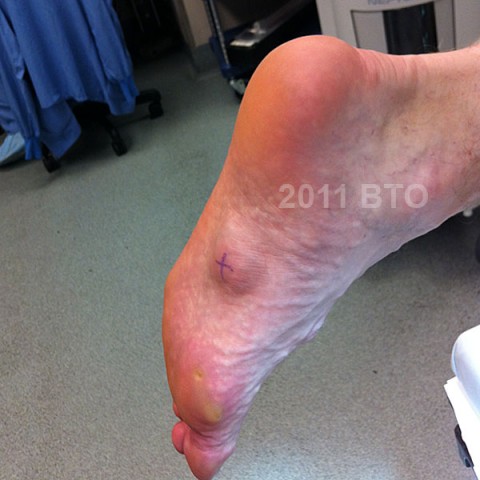
This 54 year old professional male is very active. He has had a mass on both feet for 20 years or more. The mass on the left has always been larger and is now problematic because of pain and swelling after activities.
Radiological findings:
Examination of the foot shows a substantial mass that measures 4 cm x 3.5 cm, in the middle of the instep or arch, it appears to be close to or attached to the medial band of the plantar fascia. Parts of it are very firm especially around the edges, but the center is quite compressible the area is slightly red. It is nontender. An MRI that was made in 2009 is examined. There is a mass that is contiguous with the plantar fascia, and portions of it have features consistent with plantar fascia, but other portions are essentially identical to the nearby plantar musculature. The features of this lesion do not suggest sarcoma. MRI's of both feet were made, and showed essentially identical features, except for size.
Laboratory results:
No labs were requested
Differential Diagnosis
Plantar fibroma, synovial sarcoma
Further Work Up Needed:
In a case such as this one, where bilateral, similar lesions, located in the medial part of the plantar fascia,have been slowly growing for more than 20 years, the diagnosis is fairly straightforward. In addition, if the MRIs of both feet have features entirely consistent with plantar fibroma, then a sarcoma is extremely unlikely. In this case sarcoma can be eliminated from the differential, and treatment of this lesion based on the mechanical and functional problems it presents can proceed. Successful surgical treatment of plantar fibroma requires radical fasciectomy, where the entire lesion and a sufficient margin of normal fascia and normal muscle would be entirely excised. If a more conservative surgery is performed, recurrence is virtually guaranteed.
Treatment Options:
Initial management should consist of shoe modifications and pain medication. Surgical removal is reserved for large lesions that are causing significant disability that have failed a well-documented course of non-operative care. Because recurrence is frequent following inadequate surgical removal, practitioners who are not prepared to undertake an aggressive and comprehensive resection of the lesion should not attempt to the procedure. Aggressive resection with a wide margin, as shown in the accompanying photos, is necessary to avoid recurrence, but may associated with significant complications. Although the procedure of choice has been given a term "radical fasciectomy", radical resection margins are not achieved. The actual margin achieved is typically a marginal to wide margin, where wide margins are achieved at the fascial boundaries of the lesion, but marginal margins are achieved on the skin surface of the lesion in order so that the skin may be closed without grafting, and marginal margins are achieved at the deep surface of the lesion in order to preserve the medial and lateral neurovascular bundles.
Special Features of this Case:
Surgical technique: A longitudinal incision roughly along a line between the
midpoint between the 1st and 2nd metatarsal heads and the medial
aspect of the calcaneus tuberosity is made. A large
ellipsoid portion of skin is resected in continuity with the lesion.
in order to make it possible to separate the lesion from skin at the
resected margins.
Thus, on the medial side, the dissection proceeds medial along
the arch of the foot and deep to the mass nea the knot of
Henry where the medial branch of the plantar artery and plantar nerve are located. The flexor digitorum longus is exposed at the deep
margin of the dissection. Proximally, a transverse fascial incision at least
2 full centimeters or more proximal to the most
proximal extent of the lesion is created, elevating and completing
resection the origin of the plantar aponeurosis. Dissecting deep and
lateral, the cutaneous branches of the lateral plantar
artery and nerve, are seen. Elevating the mass from proximal to distal,
the deep surface of the mass must be freed from the flexor digitorum brevis, some of which is resected en bloc along with the tumor in order to
preserve the best possible margin. The proper
digital nerves require careful dissection in order to prevent neurovascular
injury. The individual 5 or 6
separate slips of the plantar aponeurosis, are identified at their most
distal extent,. Each slip
was transected transversely at the level of the metatarsal head, several centimeters from the tumor. The tumor should be marked with a long suture on the lateral portion, and a short suture on the proximal portion, and the orientation of
the lesion and the clinical and pathological concerns must be discussed
with the pathologist.
Image
Secret Tumor Name
Image Types
Image modality
Tumor Name
Example Image
yes
Tumor Type
Benign or Malignant
Body region


