Case Identification
Case ID Number
Tumor Type
Body region
Benign or Malignant
Clinical case information
Case presentation
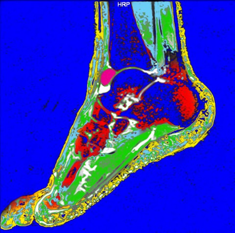
A 40 year old male presents with a mass that was found on an MRI done 17+ months ago for peroneal tendon tendonitis. A new MRI showed that the mass had gotten much larger.
Radiological findings:
A 40 year old male presents with a mass that was found on an MRI done by another provider more than 17 months previous. According to the patient, the mass was not bothersome at that time. The patient presented for a recurring issue in the same ankle. Since the lesion had not been addressed, a new MRI was ordered, which showed the mass had increased significantly in size.
Laboratory results:
No labs were done. Lab exams do not normally add useful information in these cases.
Differential Diagnosis
This is an intra-arrticular process, which means there are only a small number of possible lesions. Intra-articular synovial sarcoma and PVNS are the top two choices.
Treatment Options:
Arthroscopy was performed, and the lesion had a brown / tan pigmented appearance, making it highly likely to be PVNS. Due to the size o fht lesion, a lateral arthrotomy was made, and the lesion was completely excised with a marrginal margin.
Image
Secret Tumor Name
Case ID Number
Image Types
Image modality
Example Image
yes
Tumor Type
Benign or Malignant
Body region


