Case Identification
Case ID Number
Tumor Type
Body region
Benign or Malignant
Clinical case information
Case presentation
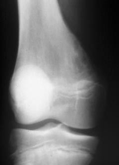
A 14 year old girl complained of a vague aching pain in her knee for two months. Exam showed a firm, slightly tender mass palpable on the surface of the posterior femur in the lateral popliteal fossa and no other findings. She had no night pain, no definite benefit from aspirin, no history of injury and was otherwise in excellent health. Laboratory exams were unremarkable.
Radiological findings:
On the plain radiographs, there is a vague sclerotic area along the posterior lateral side of the femur. The lesion is on the surface of the bone. The differential should include the list of surface lesions that occur in this location and age: Periosteal osteosarcoma, parosteal osteosarcoma, osteochondroma, osteoid osteoma, myositis ossificans, and metaphyseal fibrous defect (tug lesion).
Differential Diagnosis
The differential diagnosis should be constructed from the plain radiographs and the patient's history. Once a list of possibilities is assembled, the CT and MRI results are examined and the probability of each diagnosis is increased or decreased. The final result should be a list of two or three possibilities. Beware of the temptation to start listing possible diagnoses based on the MRI, the bone scan, or the CT, since these modalities may "overcall" the lesion.
The lesion has no aggressive features by radiographic criteria. The lesion does have a small round lucent area seen on the CT that might be interpreted as a nidus, leading to the possibility of osteoid osteoma, but the patient does not have the expected pain pattern. The reformatted CT and the lateral MRI are very helpful, since they show the how the lateral head of the gastrocnemius is inserting on the posterior femur right at the site of the lesion. This, the location, and the patient's age make the diagnosis clear.
The lesion has no aggressive features by radiographic criteria. The lesion does have a small round lucent area seen on the CT that might be interpreted as a nidus, leading to the possibility of osteoid osteoma, but the patient does not have the expected pain pattern. The reformatted CT and the lateral MRI are very helpful, since they show the how the lateral head of the gastrocnemius is inserting on the posterior femur right at the site of the lesion. This, the location, and the patient's age make the diagnosis clear.
Image
Secret Tumor Name
Case ID Number
Image Types
Image modality
Tumor Name
Benign or Malignant


