Case Identification
Case ID Number
Tumor Type
Body region
Benign or Malignant
Clinical case information
Case presentation
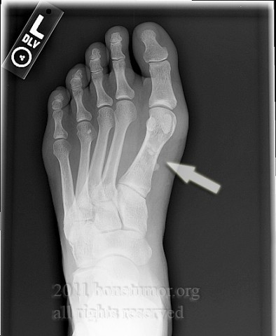
This generally healthy 36-year-old woman has calcifications in the tissues plantar to the first metatarsal. There is no history of injury.
Radiological findings:
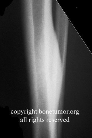
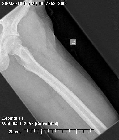
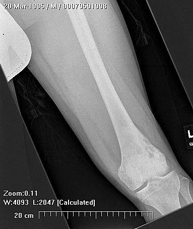
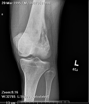
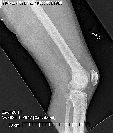
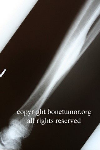
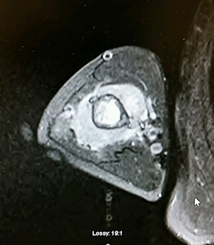
radiographs show smooth, amorphous, lobular densities or calcifications within the tissues plantar to the first metatarsal. This area corresponds to the flexor hallucis brevis. The calcifications measure 2 to 8 millimeters. MRI shows the low signal areas corresponding to the calcifications, surrounded by an area of increased signal intensity, with possible prominent vascular markings, consistent with inflammation or edema.
Laboratory results:
Do to the patient's initial presentation with redness and swelling (see photo), laboratory examination for gout and for infection was done and found to be negative. (per pt report - labs not available)
Differential Diagnosis
possibilities include heterotopic ossification, hemangioma, and less likely, synovial sarcoma.
Further Work Up Needed:
because of the clinical concern, a definitive diagnosis is necessary. Biopsy as planned.
Pathology results:
pending
Treatment Options:
if this lesion is a benign process, partial excision for diagnostic purposes only might be sufficient. Other treatments would depend on the outcome of the pathologic examination.
Special Features of this Case:
One should always be aware of the potential for synovial sarcoma. Synovial sarcoma can present with a puzzling array of very benign clinical features, and calcifications within the mass are seen in a significant proportion of these tumors. Synovial sarcoma is the most common soft tissues sarcoma in the foot, and occurs in young active healthy adults. Synovial sarcoma clearly needs to be carefully considered in this case.
Image
Secret Tumor Name
Case ID Number
Image Types
Image modality
Tumor Name
Tumor Type
Benign or Malignant