Case Identification
Case ID Number
Tumor Type
Body region
Benign or Malignant
Clinical case information
Case presentation
The patient is a healthy 25-year-old woman. She had a bump discovered near her right hip several years ago, that she thought was cellulite. It was not painful then. Recently the bump seems bigger, and it is now symptomatic.
Radiological findings:
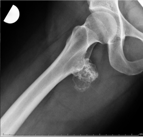
There is a pedunculated surface lesion on the posterior aspect of the right proximal femur, which measures about 50 x 40 mm in size. It projects into the muscular tissues posterior and lateral to the lesser trochanter. Portions of the lesion are densely ossified. There are some ring and arc patterns of calcification. The lesion has a cap of tissue with high signal intensity on T2 weighted images consistent with cartilage. The maximum thickness of the cap is less than 2 cm.
Laboratory results:
none requested
Differential Diagnosis
Osteochondroma versus chondrosarcoma, secondary
Further Work Up Needed:
Due to the appearance and history of this lesion, combined with the thickness of the cartilage cap, excisional biopsy appears to be a reasonable approach.
Special Features of this Case:
When a lesion that has features consistent with osteochondroma has been growing or increasing in symptoms in an adult, secondary chondrosarcoma must be considered. However, the patient's history of a bump in the area several years ago helps establish that the lesion has probably been present for some time. Large deep osteochondromas around the proximal femur may be asymptomatic until some sort of bursitis or degenerative change begins causing inflammation or pain. Patients typically will present with these in their mid-20s or even in their 30s or 40s. The symptoms are not from growth of the lesion, but rather from the tissues that are in contact with the lesion becoming symptomatic due to advancing age.
Image
Secret Tumor Name
Case ID Number
Image Types
Image modality
Tumor Name
Example Image
yes
Benign or Malignant
Body region
position within the bone


